Ed Friedlander, M.D., Pathologist
scalpel_blade@yahoo.com
No texting or chat messages, please. Ordinary e-mails are welcome.

|

|
 |
 |
 |
 |
|
verify here. |
Cyberfriends: The help you're looking for is probably here.
This website collects no information. If you e-mail me, neither your e-mail address nor any other information will ever be passed on to any third party, unless required by law.
This page was last modified January 1, 2016.
I have no sponsors and do not host paid advertisements. All external links are provided freely to sites that I believe my visitors will find helpful.
Welcome to Ed's Pathology Notes, placed here originally for the convenience of medical students at my school. You need to check the accuracy of any information, from any source, against other credible sources. I cannot diagnose or treat over the web, I cannot comment on the health care you have already received, and these notes cannot substitute for your own doctor's care. I am good at helping people find resources and answers. If you need me, send me an E-mail at scalpel_blade@yahoo.com Your confidentiality is completely respected. No texting or chat messages, please. Ordinary e-mails are welcome.
 I am active in HealthTap,
which provides free medical guidance from your cell phone.
There is also a fee site at
www.afraidtoask.com.
I am active in HealthTap,
which provides free medical guidance from your cell phone.
There is also a fee site at
www.afraidtoask.com.
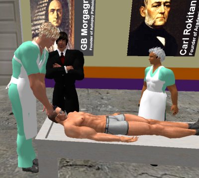
 If you have a Second Life account, please visit my teammates and me at the Medical Examiner's office. |

|
 |
With one of four large boxes of "Pathguy" replies. |
 I'm still doing my best to answer
everybody.
Sometimes I get backlogged,
sometimes my E-mail crashes, and sometimes my
literature search software crashes. If you've not heard
from me in a week, post me again. I send my most
challenging questions to the medical student pathology
interest group, minus the name, but with your E-mail
where you can receive a reply.
I'm still doing my best to answer
everybody.
Sometimes I get backlogged,
sometimes my E-mail crashes, and sometimes my
literature search software crashes. If you've not heard
from me in a week, post me again. I send my most
challenging questions to the medical student pathology
interest group, minus the name, but with your E-mail
where you can receive a reply.
Numbers in {curly braces} are from the magnificent Slice of Life videodisk. No medical student should be without access to this wonderful resource.
 I am presently adding clickable links to
images in these notes. Let me know about good online
sources in addition to these:
I am presently adding clickable links to
images in these notes. Let me know about good online
sources in addition to these:
 My team:
pathology.org -- my cyberfriends, great for current news and browsing for the general public
EnjoyPath -- a great resource for everyone, from beginning medical students to pathologists with years of experience
Medmark Pathology -- massive listing of pathology sites
Estimating the Time of Death -- computer program right on a webpage
Pathology Field Guide -- recognizing anatomic lesions, no pictures
Freely have you received, freely give. -- Matthew 10:8. My site receives an enormous amount of traffic, and I'm still handling dozens of requests for information weekly, all as a public service.
Pathology's modern founder, Rudolf Virchow M.D., left a legacy of realism and social conscience for the discipline. I am a mainstream Christian, a man of science, and a proponent of common sense and common kindness. I am an outspoken enemy of all the make-believe and bunk that interfere with peoples' health, reasonable freedom, and happiness. I talk and write straight, and without apology.
Throughout these notes, I am speaking only for myself, and not for any employer, organization, or associate.
Special thanks to my friend and colleague, Charles Wheeler M.D., pathologist and former Kansas City mayor. Thanks also to the real Patch Adams M.D., who wrote me encouragement when we were both beginning our unusual medical careers.
If you're a private individual who's enjoyed this site, and want to say, "Thank you, Ed!", then what I'd like best is a contribution to the Episcopalian home for abandoned, neglected, and abused kids in Nevada:

My home page
More of my notes
My medical students
Especially if you're looking for information on a disease with a name that you know, here are a couple of great places for you to go right now and use Medline, which will allow you to find every relevant current scientific publication. You owe it to yourself to learn to use this invaluable internet resource. Not only will you find some information immediately, but you'll have references to journal articles that you can obtain by interlibrary loan, plus the names of the world's foremost experts and their institutions.
Alternative (complementary) medicine has made real progress since my generally-unfavorable 1983 review. If you are interested in complementary medicine, then I would urge you to visit my new Alternative Medicine page. If you are looking for something on complementary medicine, please go first to the American Association of Naturopathic Physicians. And for your enjoyment... here are some of my old pathology exams for medical school undergraduates.
I cannot examine every claim that my correspondents
share with me. Sometimes the independent thinkers
prove to be correct, and paradigms shift as a result.
You also know that extraordinary claims require
extraordinary evidence. When a discovery proves to
square with the observable world, scientists make
reputations by confirming it, and corporations
are soon making profits from it. When a
decades-old claim by a "persecuted genius"
finds no acceptance from mainstream science,
it probably failed some basic experimental tests designed
to eliminate self-deception. If you ask me about
something like this, I will simply invite you to
do some tests yourself, perhaps as a high-school
science project. Who knows? Perhaps
it'll be you who makes the next great discovery!
Our world is full of people who have found peace, fulfillment, and friendship
by suspending their own reasoning and
simply accepting a single authority that seems wise and good.
I've learned that they leave the movements when, and only when, they
discover they have been maliciously deceived.
In the meantime, nothing that I can say or do will
convince such people that I am a decent human being. I no longer
answer my crank mail.
This site is my hobby, and I do not accept donations, though I appreciate those who have offered to help.
During the eighteen years my site has been online, it's proved to be one of the most popular of all internet sites for undergraduate physician and allied-health education. It is so well-known that I'm not worried about borrowers. I never refuse requests from colleagues for permission to adapt or duplicate it for their own courses... and many do. So, fellow-teachers, help yourselves. Don't sell it for a profit, don't use it for a bad purpose, and at some time in your course, mention me as author and William Carey as my institution. Drop me a note about your successes. And special thanks to everyone who's helped and encouraged me, and especially the people at William Carey for making it still possible, and my teaching assistants over the years.
Whatever you're looking for on the web, I hope you find it, here or elsewhere. Health and friendship!

![]()
![]()
If nature had wanted you to be a specialist, she'd have had you born with one eye with a microscope fastened to it.
-- Buckminster Fuller
An eye for an eye" makes the world blind.
-- Mohandas Gandhi
"The eye altering alters all."
-- William Blake
 Young men's love lies not in their hearts but in their eyes.
τυφλάς ΄εν αύτοίς ΄ελπίδας καταώικισα
I caused to dwell within human beings hopes for things that they could not see.
-- Aeschylus's Prometheus
Heaven wheels above you displaying to you her eternal glories and still your eyes are on the ground.
-- Dante
{21860} normal fundus
{21863} normal fundus
![]() KCUMB Students
KCUMB Students
"Big Robbins" -- Eye
Lectures follow Textbook
QUIZBANK
Eye & Ear #'s 1-42
|
|
|
|
|
|
|
LEARNING OBJECTIVES for this unit...
Give a reasonable differential diagnosis for conjunctivitis, uveitis, and cataracts.
Describe the abnormal anatomy and physiology of each of these, and recognize it anatomically:
INTRODUCING THE EYE
On general anatomic pathology services, pathologists seldom see material from the eye. We've
already mentioned eye disease when we talked about trachoma![]() ,
diabetes, vitamin A deficiency,
onchocerciasis (still common in much of sub-Saharan Africa: Lancet 372:
1721, 2008), and loa loa. "Big Robbins" contains a chapter on "Eye" that you will probably
want to read.
,
diabetes, vitamin A deficiency,
onchocerciasis (still common in much of sub-Saharan Africa: Lancet 372:
1721, 2008), and loa loa. "Big Robbins" contains a chapter on "Eye" that you will probably
want to read.
There are about 45 million blind people in the world, including 6 million children. It is far more common in the poor nations than in the industrial nations, and the most common causes are lack of health care, vitamin A deficiency, poor sanitation, "traditional health practices", and uncorrected refractive error. Thisis true and it is not the way that things should be. Review Br. Med. J. 327: 760, 2003.
Eye enucleations are not rare. About half are done for tumors, and a majority of the rest are taken because of trauma (Am. J. Clin. Path. 119: 594, 2003.)
* In 2007, one of the strangest events in the history of the new biotech medications began. Bevacizumab, the anti-VEGF antibody ("Avastin"), had discovered to be greatly effective when injected into the vitreous for CMV (the FDA-approved indication), the wet version of age-related macular degeneration, diabetic retinopathy, and some other eye diseases (Arch. Ophth. 126: 941, 2008; Am. J. Ophth. 146: 91, 2008). The maker of bevacizumab (Genentech) came up with a variant called Ranibizumab ("Lucentis"), which proved far more effective than any other current therapy for wet age-related macular degeneration; however, Bevacizumab, which was priced much lower, seemed to work as well. Genentech stopped preparing Bevacizumab for injection into the eye; the American Academy of Ophthalmology and other medical groups struck back and got Genentech to reverse itself. The two now seem comparable: NEJM 364: 1897, 2011.
* Future pathologists: Should you take the eyes at autopsy? The pathologists at Duke decided to examine eyes routinely at autopsies, and claim that there was something important in 32%, and at least one diagnosis in 86%. I am not sure that everyone will agree with their conclusion that eyes should always be taken, and I doubt families will always want this. See Arch. Path. Lab. Med. 125: 1193, 2001. By contrast, the pathologists at Leeds found only one surpise (the blindness was due to metastatic adenocarcinoma) in twenty years' worth of enuclated eyes on the living (J. Clin. Path. 59: 153, 2006.)
 When I was a kid, there was no routine screening |
SELECTED EYE WORDS
The following terms are the ones I wish I'd learned in my basic sciences years. Additional terms appear in the remainder of the handout.
AMAUROSIS: An eye that appears normal, but is blind. Perhaps the lesion is in the optic nerve or brain.
AMAUROSIS FUGAX: An episode of blindness lasting less than ten minutes. A variety of causes are known, including small thrombi, atheroemboli (Neurology 62: 117, 2004), temporal arteritis, and vasospasm (the last responds to calcium channel blockers NEJM 329: 396, 1993).
AMBLYOPIA: An eye that appears normal, yet cannot see normally. One important cause is the "lazy eye", i.e., the one ignored by strabismus patients.
ANGLE: The angle in the anterior chamber where the uveal tract joins the cornea-sclera. This is where the aqueous humor drains from the eye.
APHAKIA: A birth defect in which the child has no lens. Modern treatment: Arch. Ophth. 128: 21, 2010.
ARCUS: Fatty deposit around the limbus of the cornea. "Arcus lipoides". Don't give it much attention; these people's LDL's are more likely to be high and their HDL's to be low (Neth. J. Med. 55: 184, 1999; Am. J. Ophth. 137: 363, 2004; no real independent indicator of risk BMJ 343: d5497, 2011). A young person with arcus can have high LDL's, have a corneal dystrophy, or be normal. We prefer not to say "arcus senilis".
BUPHTHALMOS: Swollen globe, as in childhood glaucoma.
CATARACT: Any opacification of the crystalline lens
COLOBOMA: A deformation of the iris / ciliary body from failure of closure of a choroid fisssure. The classic coloboma looks like a classic keyhole.
{22238} coloboma
COLOR BLINDNESS: Inherited defects in one or more of the cones. Most are X-linked recessives. Common red-green blindness is well-known; deuterans have trouble seeing blue and green, protans have trouble seeing red, etc.
COTTON-WOOL PATCH: An ischemic area of the nerve-fiber layer of the retina
CYCLITIS: Inflammation of the ciliary body
DACROCYSTITIS: Inflammation of the lacrimal apparatus
{13179} dacryocystitis
DOT-AND-BLOT HEMORRHAGES: Bleeding into the inner nuclear layer of the retina
DRUSEN: Accumulation of inert material (described as a mix of protein and lipofuscin) at the basement membrane of the choroid.
ECTROPION: Eversion of an eyelid
ENDOPHTHALMITIS: Inflammation of the interior of the eye
ENTROPION: In-turning of an eyelid, often as a result of scar contraction on its conjunctival surface. This sounds bad and is. Often the eyelashes scratch the cornea (TRICHIASIS).
EPICANTHUS / EPICANTHIC FOLD: An extra fold of skin next to the nose, imparting a distinctive contour to the periorbital tissue
ESOTROPIA: Crossed eyes; convergent strabismus
EXOPHTHALMOS: When the eyeballs are pushed too far forward (usually both eyes, "proptosis" if only one)
EXOTROPIA: Wall-eyes; divergent strabismus
FLAME HEMORRHAGE: Bleeding into the nerve-fiber layer of the retina
FLUORESCEIN: Dye used in ophthalmology to better visualize breaks in the surface of the cornea.
HYPEROPIA / HYPERMETROPIA: Farsightedness, convergence of focus posterior to the retina. Often, but not always, due to a foreshortened eyeball.
HYPERTELORISM: Eyes set very wide apart.
HYPHEMA: Blood in the anterior chamber
HYPOPYON: Pus in the anterior chamber
HYPOTELORISM: Eyes set very close together
IRITIS: Inflammation of the iris
KERATIC PRECIPITATATE: Clusters of chronic inflammatory cells on the inner surface of the cornea. "Mutton-fat"; seen in uveitis.
KERATITIS: Inflammation of the cornea
KERATOCONUS: Cone-shaped cornea
{22180} keratoconus
LEUKOCORIA: White instead of the normal red pupillary light reflection. Think of retinoblastoma!
LIMBUS: The junction between the cornea and the sclera
MARCUS GUNN PUPIL: Descriptions vary, but this is an indicator for optic nerve damage in the involved eye. The pupil is larger than the other one ("to let in more light"), and constricts more slugglishly than the other on both direct and consensual testing.
MYOPIA: Nearsightedness, convergence of focus anterior to the retina. Often, but not always, due to an elongated eyeball.
PANOPHTHALMITIS: Inflammation of the whole eye. Usually bacterial, after trauma.
PAPILLEDEMA: Changes in the optic disk seen in increased intracranial pressure. Mechanisms are complex and reviewed in "Big Robbins"; longstanding papilledema damages the optic nerve.
{22092} papilledema
PHACO-, PHAKO-: Pertaining to the lens of the eye
PHOTOPHOBIA: Pain on exposure to light
PHTHISIS BULBI: End-stage, shrunken, often painful eye
PRESBYOPIA: Loss of the ability of the lens to alter its shape; one result of advancing age. The usual reason people get bifocal eyeglasses.
PROPTOSIS: Forward protrusion of the eye (usually just one eye; "exophthalmos" if it's both eyes)
RETINAL DYSPLASIA: Persistence of many little tubules and/or "rosettes", like in embryonic eye or classic retinoblastomas. Seen, usually with other abnormalities, in certain birth defects.
SCOTOMA: A blind spot
STRABISMUS: Eyes pointed in different directions, the result of eye muscle balance problems. Several interesting synonyms exist, including (for some reason) "squint". NEJM 356: 1041, 2007. Operating babies is now evidently the norm.
SYNECHIAE: As elsewhere, fibrous adhesions resulting from healing
UVEA / UVEAL TRACT: Term for the vascular coat of the eye, i.e., the choroid, ciliary body, and iris.
XEROPHTHALMIA: Dry eyes
BIRTH DEFECTS: A selection
ANOPHTHALMOS / ANOPHTHALMIA (i.e., no ocular tissue in the orbit) is rare and seen only with extreme malformations of the brain.
MICROPHTHALMIA, one or both eyes too small, has a variety of causes, including intrauterine infection (CMV, rubella, toxoplasmosis), fetal alcohol syndrome, trisomy 13 or 18, or any of a variety of genetic syndromes (173 at last count: Am. J. Ob. Gyn. 194: 1354, 2006).
TRISOMY 13 ("Trisomy D", "Patau's")
You're already familiar with the other stigmata of this illness. The eyes are almost never normal. They may be single ("cyclops"; perhaps only a retina) or fused (cyclops variant); even if there are the correct number, they present an array of abnormalities described in "Big Robbins".
* Future eye pathologists: A coloboma with cartilage suggests Patau's.
TRISOMY 21 ("Down's", etc.)
BRUSHFIELD'S SPOTS are areas where the iris is hypoplastic.
{15608} Brushfield's spots
A variety of other abnormalities are common. Remember esotropia and epicanthic folds.
CONGENITAL RUBELLA SYNDROME![]() syndrome
syndrome
Again, you are familiar with the other features of this syndrome.
Rubella![]() cataract results from retention of nuclei in the center of the lens. This is pretty distinctive.
cataract results from retention of nuclei in the center of the lens. This is pretty distinctive.
* The iris epithelium is largely necrotic, there may be granulomas, and so forth. The result is LEATHER IRIS.
* The pigment layer of the retina alternates between hypertrophic ("pepper") and atrophic ("salt") areas.
* Congenital rubella![]() syndrome with blindness is still rampant
where immunization is not practiced, or where people refuse (Arch. Ophth. 122: 541, 2004;
J. Inf. Dis. 187(S1): S-146 & S-191 & S-223 & S-235, 2003; Am. J. Pub. Health 90: 1555, 2000).
The danger period is the first three months of pregnancy.
syndrome with blindness is still rampant
where immunization is not practiced, or where people refuse (Arch. Ophth. 122: 541, 2004;
J. Inf. Dis. 187(S1): S-146 & S-191 & S-223 & S-235, 2003; Am. J. Pub. Health 90: 1555, 2000).
The danger period is the first three months of pregnancy.
Rubella remains rampant in the poor nations, where it kills maybe half a million babies yearly. During the 1990's, the anti-immunization movement resulted in the birth of 58 children with congenital rubella in the USA.
CONGENITAL SYPHILIS![]()
Again, you are familiar with the extra-ocular features.
The characteristic eye finding is inflammation of corneal stroma (INTERSTITIAL KERATITIS). This appears in youth, but is not present at birth.
There are likely to be other abnormalities in the eyes.
TAY-SACH'S
In this and a few other inborn errors of metabolism, the normal red of the macula forms the "cherry" in the pale, lipid-laden retina.
{20115} cherry red spot
LID PROBLEMS
STY (HORDEOLUM) is suppuration (usually
staphylococcal![]() )
of the hair follicle, the sebaceous glands of the eyelash
(* "Zeis's glands"), and/or apocrine glands (* "Moll's glands").
)
of the hair follicle, the sebaceous glands of the eyelash
(* "Zeis's glands"), and/or apocrine glands (* "Moll's glands").
![]() CHALAZION (* Greek for hailstone) is chronic, usually granulomatous, inflammation of the large sebaceous glands
(* "Meibomian") glands of the eyelid that produce the film of oil that keeps tears from evaporating.
CHALAZION (* Greek for hailstone) is chronic, usually granulomatous, inflammation of the large sebaceous glands
(* "Meibomian") glands of the eyelid that produce the film of oil that keeps tears from evaporating.
{13143} chalazion
Of course, infections that involve the bulbar conjunctiva often involve the conjunctiva of the lid, too. See below.
VITAMIN A DEFICIENCY (Br. Med. J. 310: 1051, 1995; Am. J. Clin. Nutr. 73: 1045, 2001)
The basic problem (at the front of the eye) is excessive squamous differentiation of the corneal epithelium ("xerophthalmia"). BITOT'S SPOTS are actually just the thickened, hyperkeratotic epithelium, typically on the conjunctiva just outside the limbus.
As you'd also expect, the corneal mucus cells are lost. Hence, the tears dry too fast.
When KERATOMALACIA is said to be present, it means the hyperkeratotic epithelium has become soft. It may become secondarily infected (* Corynebacterium xerosis, others) and even perforate.
Remember that in the absence of vitamin A, you also can't make the pigment for rods (at the back of the eye). Night blindness results.
Many governments, for their own dark reasons, fail to implement or even allow public health measures that would do a lot of good for very little money spent. Over half of the boys in rural Ethiopia have Bitot's spots (East African Medical Journal 76: 590, 1999); of course their growth is stunted and their vitamin A levels very low. Cambodia: Arch. Ophth. 122: 517, 2004.
An amateur vegan family blinds its six-year-old son: Arch. Ophth. 122: 1228, 2004. More about inept vegans: Clin. Ped. 43: 107, 2004.
* After years of delay because of anti-biotech activism, vitamin-A enriched rice is now widely used and being welcomed by the world's poor: Nature Biotech. 21: 971, 2003.
TRACHOMA (Br. Med. J. 362: 223, 2001; Lancet 362: 223, 2003; NEJM 358: 1777, 2008; Lancet 371: 1945, 2008; Lancet 373: 1111, 2009)
We've already discussed Chlamydia trachomatis![]() ,
an important cause of blindness worldwide. Remember that the serovars that
cause trachoma and those that cause genital infection are distinct.
,
an important cause of blindness worldwide. Remember that the serovars that
cause trachoma and those that cause genital infection are distinct.
The pathology mostly involves lymphoid follicles on the conjunctival membrane. Later there is scarring.
If there is coexistent vitamin A deficiency, the infection is likely to be worse (Lancet 357: 1676, 2001).
McCallan's stages of trachoma:
I: Follicles (i.e., lymphoid infiltrates) on the cornea, with early fibrosis
II: Macrophages laden with the debris of dead human cells ("Leber cells") appear
III: Severe scarring; contraction produces an entropion
IV: The actual infection self-cures, since the damaged, opacified eye surface is no longer vulnerable to infection.
* For that matter, simply building latrines that people can use seems to reduce the prevalence of trachoma (Am. J. Trop. Med. 82: 693, 2010).
CONJUNCTIVITIS (JAMA 310: 1721, 2013)
Trachoma's![]() lesser
chlamydial counterpart in the U.S. is SWIMMING POOL CONJUNCTIVITIS / INCLUSION
BODY CONJUNCTIVITIS, generally from genital chlamydia rather than the
endemic trachoma microbes. A baby may catch chlamydia from Mom while being born.
Look for the characteristic inclusions on smears (* "elementary bodies"; we suggest a Giemsa stain).
lesser
chlamydial counterpart in the U.S. is SWIMMING POOL CONJUNCTIVITIS / INCLUSION
BODY CONJUNCTIVITIS, generally from genital chlamydia rather than the
endemic trachoma microbes. A baby may catch chlamydia from Mom while being born.
Look for the characteristic inclusions on smears (* "elementary bodies"; we suggest a Giemsa stain).
BACTERIAL CONJUNCTIVITIS generally produces a red eye with purulent discharge. Think of gonorrhea,
pneumococci, staph![]() (* I once endured this), and Hemophilus.
(* I once endured this), and Hemophilus.
Classic OPHTHALMIA NEONATORUM was gonococcal infection acquired in the birth canal. Silver nitrate, erythromycin, and (now) povidone iodine (great for the poor nations) are prophylaxis (NEJM 332: 562 & 600, 1995).
Conjunctivitis can also be viral (remember adenovirus![]() which used to travel on ophthalmologists' instruments,
and measles
which used to travel on ophthalmologists' instruments,
and measles![]() ), IgE-mediated ("vernal
conjunctivitis", i.e., in the springtime allergy season; look for eosinophils on smear), due to delayed
hypersensitivity (often cosmetics), due to injury or a foreign body, or due to ultraviolet light
(remember "snow blindness").
), IgE-mediated ("vernal
conjunctivitis", i.e., in the springtime allergy season; look for eosinophils on smear), due to delayed
hypersensitivity (often cosmetics), due to injury or a foreign body, or due to ultraviolet light
(remember "snow blindness").
Remember that conjunctivitis from most causes can involve the cornea (KERATOCONJUNCTIVITIS). See below. Unless you know the cause of the conjunctivitis, don't share towels....
|
|
PINGUECULA AND PTERYGIUM
PINGUECULA is fatty deposits in the conjunctiva, especially in people who've had lots of sun exposure. Look near the nasal aspect of the limbus on both sides.
{05932} pinguecula
PTERYGIUM ("wing") is more of the same, but extending over the cornea, obliterating Bowman's membrane.
{21874} pterygium, gross
{21876} pterygium, gross
{21879} pterygium, micro, oil red O stain for lipid
CORNEAL DISEASE
Remember the layers (from out to in): Epithelium, Bowman's membrane, stroma, Descemet's membrane, endothelium.
Because there are no vessels here, once the cornea gets infected, it's serious. Especially in parts of the world where treatment is not available, blindness often results.
HERPES SIMPLEX![]() produces the familiar dendritic ulcers.
produces the familiar dendritic ulcers.
{14127} herpes keratitis
{21931} herpes keratitis
{22055} herpes, inclusion bodies![]()
ACANTHAMOEBA produced an epidemic among users of certain contact-lens solutions in 1986 (MMWR June 27, 1986), and there are occasionally voluntary recalls of solutions that may be contaminated.
{22192} red eye from acanthamoeba
|
|
Kayser-Fleischer ring: copper deposit in Wilson's disease. See it, and it's probably too late for your patient.
Band keratopathy: Hypercalcemia. See it, and you've probably been missing hypercalcemia for way too long.
{21992} Kayser-Fleischer ring
{21927} band keratopathy, gross
{21928} band keratopathy, micro, calcium dark blue
* The CORNEAL DYSTROPHIES, which render the cornea opalescent due to faulty chemistry, include several hereditary problems.
Granular dystrophy -- autosomal dominant -- opacities
Lattice dystrophy -- autosomal dominant -- amyloid
Macular dystrophy -- autosomal recessive -- mucopolysaccharide
Also worth knowing: Fuch's -- leaky backside epithelium, with water bubbles on the inner surface
Here's an example of one locus for several illnesses: BIGH3 can cause lattice, granular, or other corneal dystrophies depending on the mutation (Am. J. Hum. Genet. 62: 320, 1998). Why is this not surprising, especially in the cornea?
{21935} granular dystrophy
{21938} granular dystrophy
{21920} lattice dystrophy
{21957} lattice dystrophy, congo red
{21929} macular dystrophy, histology
{21932} macular dystrophy, histology
{22117} corneal transplant
{22121} corneal transplant
If for some reason you wish to know more about amyloid on the cornea and elsewhere in the eye, refer to Am. J. Ophth. 117: 529, 1994.
UVEAL DISEASE
ANIRIDIA: No iris. Lots of problems with the eyes; many of these patients have a gene deletion that extends to the nearby Wilms tumor locus (chromosome 11).
ALBINISM: The major problem these people face is excessive sensitivity of the eye to light.
NON-GRANULOMATOUS UVEITIS is usually idiopathic, chronic inflammation.
GRANULOMATOUS UVEITIS (by contrast) has a long differential...
Infection
Bacteria: tularemia![]() ,
syphilis
,
syphilis![]() , gonorrhea
, gonorrhea
Viruses: CMV![]() ,
zoster
,
zoster![]() (remember CMV retinitis as well)
(remember CMV retinitis as well)
Fungi: various
Other: toxoplasmosis![]() .
(NOTE: Toxoplasmosis is an important disease of the eye. In the worst
cases, pathologists find patches of
coagulation necrosis
.
(NOTE: Toxoplasmosis is an important disease of the eye. In the worst
cases, pathologists find patches of
coagulation necrosis![]() in retina and choroid.)
in retina and choroid.)
Odd immune diseases
Sarcoidosis
Ulcerative colitis and Crohn's disease
Reactive arthritis ("Reiter's") and Behçet's syndromes
Juvenile rheumatoid arthritis (infamous; don't overlook this one as treatment can really help prevent loss of vision: Am. J. Ophth. 143: 840, 2007).
SYMPATHETIC OPHTHALMIA is probably an autoimmune reaction against one's own pigment epithelium (iris and choroid) and (to a lesser extent) retinal neurons following penetrating injury of one eye. Unfortunately, it involves both eyes, and does great harm. Thankfully it is rare, but the only sure prevention is to enucleate the injured eye within a week or two. A white-knuckle management problem for ophthalmologists. The anatomic pathology: Am. J. Ophth. 138: 475, 2004.
Most uveitis that will lead to permanent vision loss is idiopathic.
* One group claims to have found evidence of infection by fastidious bacteria from about half of their large series of uveitis patients (Medicine 87: 167, 2008); the microbes include chlamydia, spirochetes, bartonella, coxiella, Whipple bacillus, and some even more obscure entities. The group emphasizes that the significance of the findings is unclear, and whether we will be sampling vitreous and sending it for "PCR for everything" is unknown.
|
|
CATARACTS
A variety of opacities in the lens; NUCLEAR SCLEROSIS is the most common type seen in the aging lens.
{12428} cataract
{22009} cataract
{22209} cataract
{22212} cataract
{22251} cataract
{22254} cataract; nuclear sclerosis
Remember as causes of cataract:
While we're on the subject of lens disease, a displaced lens ("ectopia lentis") is one problem faced by Marfan types.
{21999} ectopia lentis
RETROLENTAL FIBROPLASIA ("retinopathy of prematurity"; Ped. Clin. N.A. 50: 77, 2003)
The developing fetal retinal circulation, especially at its temporal margin, is selectively damaged by high concentrations of oxygen. The vessels, which are sprouting from the optic disk, at first constrict, then obliterate, so that the peripheral retina fails to vascularize. Several weeks after cessation of oxygen therapy, vessels begin sprouting willy-nilly from the edge, and grow into the vitreous; scar may contract and detach the retina, and there may be other problems.
This is a classic cause of blindness in premature infants treated with high concentrations of oxygen. It became a major public health problem in the 1940's and early 1950's (my grammar school had a girl a few years older than myself who had a full-time amanuensis). It became less troublesome when oxygen concentrations were reduced, but has recurred now that very small babies are surviving (a 600 gm baby tolerates even 90 mmHg of oxygen poorly).
Does attempting to keep oxygen saturations at 85-89 mmHg rather than 91-95 mmHg led to an increased risk of death? Yes! NEJM 368: 2094, 2013. No! JAMA 309: 2111, 2013.
Laser treatment is now the mainstay of treatment, and is a great help even in the poorest communities (Am. J. Ophth. 154: 750, 2012).
HYPERTENSIVE AND ARTERIOSCLEROTIC (better, "arteriolosclerotic") RETINOPATHY (Lancet 369: 425, 2007).
A common subject for discussion on rounds. Here's an old system that's passing out of use.
HYPERTENSIVE RETINOPATHY: High blood pressure is bad for the vessels.
Grade I: The arterioles are a bit narrowed (do you think this is a cause or an effect of hypertension? nobody knows....)
Grade II: The arterioles are going into spasm somewhere, i.e., noticeably narrower than the veins
Grade III: There are bleeds and/or cotton wool patches (ischemic areas)
Grade IV: There is also edema of the optic disk, i.e., the hypertension has resulted in increased intracranial pressure
{22018} hypertensive retinopathy with hemorrhages
ARTERIO(LO)SCLEROTIC RETINOPATHY: Due to progressive opacification of the arteries
Grade I: Widened light reflex due to "hyaline arteriolosclerosis", which renders the vessels opalescent
Grade II: AV crossing defects; the hyaline wall of the artery makes it hard to see the vein underneath ("arterial sheathing" showcases the thickened translucent wall of the artery against the vein; "arterial nicking" is the extra-thick artery pressing on the vein)
Grade III: Copper wires; the blood doesn't show clearly through the arterial wall, making it look orange
Grade IV: Silver wires. The blood fails to show at all.
{22034} don't forget diabetic retinopathy, too
RETINITIS PIGMENTOSA
A heterogeneous group of diseases, mostly hereditary, in which the photoreceptor cells gradually die off. Patients are first troubled by loss of peripheral vision, and later lose all sight. The rods are typically lost before the cones ("night blindness", again).
There is proliferation of the pigment cells of the retina, typically along the vessels.
The molecular biology for the major autosomal dominant forms were worked out in the early 1990's. Around 1/3 of cases have a defective rhodopsin gene; some people with simple autosomal-dominant night-blindness have the same thing (Nature 367: 639, 1994), and vitamin A in fairly big doses can help these folks.
* Other autosomal dominant syndromes are known (RP1, peripherin, NRL: Br. J. Ophth. 86: 328, 2002; KLHL7 Arch. Ophth. 128: 772, 2010).
In common (rhodopsin) retinitis pigmentosa, the defective rhodopsin accumulates and kills the cells. Gene therapy works in mice, and involves administering a ribozyme (via a virus) that cleaves the defective gene's mRNA (Nat. Med. 4: 967, 1998.)
* Usher syndrome (retinitis pigmentosa and congenital nerve deafness) is often the myosin VII-A locus. The newly-discovered BBS1 gene has a variety of alleles that produce retinitis pigmentosa, either alone or with Bardet-Biedl (Arch. Ophth. 130: 1425, 2012).
Consider night blindness in children with unexplained fear of the dark.
{22074} retinitis pigmentosa
![]() Retinitis pigmentosa
Retinitis pigmentosa
Prize photograph
Institute of Medical Illustrators
STATIONARY NIGHT BLINDNESS, i.e., the rods don't work but the cones work fine, has a variety of genetic causes (Nat. Genet. 13: 358, 1996; update Am. J. Ophth. 135: 733, 2003).
Vitamin A deficiency is of course a better-known cause of night blindness, but thankfully it is no longer common in the U.S. except among people on crackpot diets.
Night-vision goggles are now coming into use for people with night-blindness (Inv. Op. 45: 1725, 2004).
RETINAL DETACHMENT
When the nerve-cell layers separate from the pigmentary epithelium, they lose their effective blood supply, and degenerate over days or weeks.
Underlying causes are (1) contraction of scar within the vitreous (as after trauma, hemorrhage); (2) exudation from inflamed or neoplastic tissue; (3) a hole in the retina, through which the vitreous penetrates.
Important causes of retinal detachment include trauma, diabetes (from neovascularization of vitreous), and marked nearsightedness (in which the eyeball is elongated and the retina stretched).
{22080} retinal detachment
{22802} retinal detachment
MACULAR DEGENERATION (Am. J. Med. 121: 279, 2008; Lancet 379: 1728, 2012)
A family of diseases in which the nervous tissue of the macula degenerates, causing lost of central vision.
The most troublesome is the common AGE-RELATED MACULAR DEGENEREATION (NEJM 358: 2378, 2008; again, it's better not to call it "senile"....) The histopathology is mild inflammation and eventualy necrosis and fibrosis (with a lot of lipid) involving the retina. Look for drusen around the macula, and maybe some new vessels in the choroid. Smoking clearly brings it on faster. Whether sunlight exposure is really a risk factor is far from clear ("maybe yes": Arch. Ophth. 119: 246, 2001; "no": Arch. Ophth. 119: 1463, 2001; "actually less if there's been heavy sun exposure" Ophth. 104: 770, 1997). Having blue eyes is at best a minor risk (Ophth. 105: 1359, 1998). High HDL (which is good for your arteries) also seems to be a risk facator (Arch. Ophth. 128: 750, 2010). Possibly zinc and anti-oxidants slows it down, vitamins C and E and carotene flopped ("Age-Related Eye Disease Study", Arch. Ophth. 119: 1417 & 1439, 2001); a more recent study trying to relate diet to macular degeneration didn't show anything striking (Arch. Ophth. 125: 1225, 2007). In smokers, is it perhaps cadmium toxicity (Mayo -- Am. J. Ophth. 144: 414, 2007)????
The most common gene for macular degeneration has been cloned, and it is a forme fruste of a young-onset disease (* Stargardt's; gene is ABCA4; Science 277: 1765, 1997). Several others are known: NEJM 351: 346, 2004. Protective allele and the possibile gene therapy target: NEJM 359: 1456, 2008.
The large majority of these patients have the "dry / geographic atrophy" variant. About 10% of patients with age-related macular degeneration have the "wet" form, with considerable growth of new blood vessels. This is the kind most likely to cause severe visual impairment, and was at the center of the "Lucentis" flap of 2007-8. These patients are now have vision stabilized and even improved from injected anti-VEGF.
* The mouse model, induced by autosensitization, features inflammation and complement deposition in Bruch's membrane (Nat. Med. 14: 194, 2008).
* The implantable telescope for end-stage macular degeneration: Am. J. Ophth. 146: 664, 2008.
The first apparently-successful use of human EMBRYONIC stem cell is the now-ongoing trials for Stargardt's and dry macular degeneration (Lancet 379: 713, 2012).
Follow-up: The embryonic stem cell patients are doing well at up to 37 months: Lancet 385: 509, 2015.
{22059} macular degeneration
{22062} macular degeneration
OPTIC NERVE
OPTIC NEURITIS has come to include any non-neoplastic cause of optic nerve dysfunction, from methanol poisoning to ischemia to demyelination (this may herald multiple sclerosis).
DEVIC'S DISEASE includes full-thickness demyelination of an optic nerve. We reviewed this under "multiple sclerosis", from which it is distinguished by the presence of "NMO-Ig" antibodies against aquaporin.
LEBER'S HEREDITARY OPTIC ATROPHY is a group of inherited diseases, one of which was the first shown to be caused by a mitochondrial gene mutation. Gene therapy helps some, temporarily, for a different Leber's variant: NEJM 358: 2240, 2008; update Lancet 374: 1597, 2009; update NEJM 372: 1887 & 1920, 2015.
GLAUCOMA (Lancet 377: 1367, 2011)
A family of diseases, their common feature being damage to the eye due to increased intraocular pressure.
(PRIMARY) OPEN-ANGLE GLAUCOMA (NEJM 360: 1113, 2009): Some problem, more or less mysterious, exists with drainage of aqueous humor via the canal of Schlemm, despite apparently normal anatomy.
* The most important gene is TIGR/MYOC/GLC1A, myocilin), a trabecular meshwork protein that in its mutated form gums up the slits: Science 275: 668, 1997; NEJM 338; 1022, 1998; another gene optineurin Science 295: 1077, 2002 & Am. J. Path. 169: 1976, 2006). CDKN2B-AS1 Am. J. Ophth. 155: 342, 2013.
CLOSED-ANGLE GLAUCOMA: Some people's anterior chamber is too shallow. As a result, the angle is too acute, and the drainage of aqueous humor is compromised. This is exacerbated as the lens thickens during later life.
CONGENITAL GLAUCOMA: Autosomal recessive (* a cytochrome C gene
component: Am. J. Ophth. 131: 345, 2001; update Am. J. Ophth. 142:
993, 2006), or rubella![]() .
.
SECONDARY GLAUCOMA, the result of some other disease or injury to the eye, may be open-angle or closed-angle.
The optic cup becomes wider and deeper, and the optic nerve and retina bear the brunt of the troubles. "Big Robbins" suggests that the increased pressure causes disruption of the axoplasmic flow where the axons lie over the edge of the deepened optic cup.
{21990} optic nerve, glaucoma
{21993} optic nerve, glaucoma, micro
EYE TUMORS
Cancer of the CONJUNTIVA AND LIDS is usually squamous cell carcinoma. As with squamous cell carcinoma of the skin, sunlight is a risk factor. The in situ phase is likely to appear as leukoplakia. Remember melanomas can occur on the outer surface of the eye; they may be preceded by "melanosis".
By far the most common eye tumor in an adult is metastatic carcinoma.
{21900} squamous cell carcinoma
{21901} squamous cell carcinoma
{21894} carcinoma in situ, gross
{21898} carcinoma in situ, gross
{21895} carcinoma in situ, micro
{21899} squamous cell carcinoma, microscopic (good pearls)
{19377} amelanotic melanoma of the conjunctiva
|
|
|
![]() Melanoma of the eye
Melanoma of the eye
AFIP
Wikimedia Commons
OCULAR MELANOMA (Arch. Path. Lab. Med. 134: 1778, 2010), from the uveal melanocytes (not the pigment epithelium) is the most important adult eye cancer.
Eye melanomas have their own common genetic signature (NEJM 363: 2191, 2010). They range from masses on the iris (often amelanotic; these tend to stay localized) to lesions hidden deep within the eyeball.
There are no lymphatics in the eye. Eye melanomas metastasize to the liver, often apperaing a decade or two after removal of the primary, accounting for the old adage "Beware the patient with the glass eye and the large liver".
There is a tremendous amount written about the genetics and histopathology of choroidal melanomas; the only thing you'll need for roundsmanship is "spindle cells are good."
You may see "plaque radiotherapy" (isotope-containing needles are placed on the eye surface over the tumor) used to save there people's vision (Am. J. Ophth. 149: 70, 2010). Gamma knife surgery for today's eye melanoma patient: J. Neurosurg. 117-S: 108, 2012.
{22162} melanoma
{22157} melanoma
{22636} melanoma
{22644} melanoma
{21873} melanoma
{21917} melanoma
{21918} melanoma
RETINOBLASTOMA (Lancet 379: 1436, 2012; Arch. Path. Lab. Med. 133: 1203, 2009; Arch. Path. Lab. Med. 136: 190, 2012) accounts for 1% of pediatric cancer deaths. There are only about 9000 worldwide but in the poor nations, most patients die.
Remember the Rb anti-oncogene? Familial cases are typically multifocal (why?), and other tumors (notably osteosarcomas) are also common in survivors, since many of these patients have been radiated, and many have germline deleted Rb.
Histopathology: Arch. Path. Lab. Med. 130: 1669, 2006. Since children almost never die any more as a result of retinoblastoma, the grading system (the fewer rosettes, the higher the grade, and necrosis is uncommon but bad) is a guide to treatment.
The extent of invasion, as seen in the enucleated eye, is a determiner of prognosis, but beyond this, the pathology team has little to offer for prognostication (Arch. Path. Lab. Med. 133: 1199 & 1210, 2009).
* Old talk about "spontaneous maturation" of retinoblastomas -- which is common in neuroblastomas -- probably just reflects the presence of residual benign retinomas / retinocytomas, the tough little benign tumors that mimic photoreceptors and from which retinoblastomas arise.
{22149} retinoblastoma, gross
{22141} retinoblastoma, gross
{22146} retinoblastoma, micro
|
|
BASAL CELL CARCINOMA of the eyelids is classic. Optic nerve MENINGIOMA, and OPTIC NERVE GLIOMA (astrocytoma -- think of neurofibromatosis; see Neurology 63: 1944, 2004) within the optic nerve, are additional eye tumors. SEBACEOUS CARCINOMA of the Meibomian glands is a rarity. Other important orbital tumors include LYMPHOMAS in adults and EMBRYONAL RHABDOMYOSARCOMA in children.
* The famous "Phillips case" of 1961 involved a Linda Epping,
an eight-year-old
girl with embryonal rhabdomyosarcoma of the eye.
The mother refused surgery in favor of being treated
by Marvin M. Phillips, a chiropractor who had supposedly "cured" a similar lesion
by nutrition and manipulation. The chiropractor promised a cure.
The cancer progressed rapidly, the child died, even the other
chiropractors testified against him, and the chiropractor
was convicted of second-degree murder.
This was a landmark case, since it was the first
conviction of murder-by-quackery.
Click here for a court opinion.
The chiropractor finally got the murder
conviction overturned by the California Supreme Court after he
had served three-and-a-half years of his sentence. I'm not a lawyer,
but the question was whether killing a child this was similar to other
felony murders, i.e., somebody dies without your intending it because you're committing a felony.
Here the felony was grand larceny for taking the parents' money on false pretenses.
Today's "reform" chiropractors look to the "Phillips case" as a landmark
that helped clean up the profession.
![]() Basal cell carcinoma
Basal cell carcinoma
Eye pathology site
{21862} sebaceous carcinoma
{21864} sebaceous carcinoma
{21865} sebaceous carcinoma, oil-red O stain (sebum)
{22101} optic nerve glioma (astrocytoma)
{22113} rhabdomyosarcoma
{22114} rhabdomyosarcoma
{22115} rhabdomyosarcoma
| * Blinding of enemies, individually or en masse, is an unpleasant fact of history. They would be removed either with sharp instrument or red-hot iron. Review: Doc. Ophth. 74: 119, 1990. Future major ground wars may make use of weapons that blind the enemy soldiers by combat troops or snipers. The weapons are already in existence and work well. They were banned by the "Protocol on Blinding Laser Weapons" treaty of 1980. Blinded soldiers require more care than dead people, and blinding is demoralizing to military and civilians alike (Br. Med. J. 315: 1392, 1997). You can get a powerful laser that looks just like a classroom pointer on the internet but can burst balloons and blind you (NEJM 363: 1089, 2010). In lawless eras, blinding one's political enemies and prisoners of war (sometimes en masse) was fairly common, and was considered a humane alternative to simply killing them. Of course, sending thousands of blinded soldiers back to their homes is worse for the enemy's economy, too. |  Rembrandt, "The Blinding of Samson" |
* In the African kleptocracies, many children are confined to "charitable institutions for the blind" simply because they are myopic and do not have glasses (Med. J. Aust. 167: 351, 1997). I leave you to figure out the dark and corrupt politics responsible for this travesty.
* Future pathologists: When the heart stops, intraocular pressure (which largely results from arterial pressure) drops by about half, and pressure continues to drop over the following hours, imparting the softness and deformability to the eyes of the dead.
| * For your reading enjoyment: Blindness is a perennial symbol for spiritual darkness. See Oedipus the King, and the Christian scriptures ("Light of the body", "Blind leading the blind"), Shakespeare's King Lear, Goethe's Faust (at the end), Robert Lewis Stevenson's Treasure Island (Blind Pew), Yeats's "On Baile's Strand" & "The Cat and the Moon", hymn "Amazing Grace", "The Blind Men and the Elephant" (re-read that last verse), cartoon "Mr. Magoo's Christmas Carol" (Scrooge isn't a bad man, he's a blind man), others. For the opposite view, see John Milton's sonnet "On His Blindness", and Paradise Lost Book III vv. 1 ff.; Ibsen's Solveig ("Peer Gynt") is blind to Peer's faults, which is how Peer eventually discovers his "real self"; the Mountain King ("In the Hall of the") offers Peer eye surgery so that he will see ugly ("Be selfish") as beautiful ("Be yourself"). Jose Saramago's "Blindness" (1995, there's a movie) might be the most famous novel from Portugal. |

|
|
|

Goya, "The Blind Guitarist"
*SLICE OF LIFE REVIEW
1021-1040, 1042-1044 retina
{15065} eye, human
{15066} eye, human
{15067} iris and lens, normal
{15067} iris and lens, normal
{15068} iris and lens, normal
{15068} iris and lens, normal
{15069} ciliary body, normal
{15069} ciliary body, normal
{15070} ciliary body, normal
{15070} ciliary body, normal
{15071} retina (cross section), normal
{15071} retina (cross section), normal
{15072} retina (cross section), normal
{15072} retina (cross section), normal
{15073} photoreceptor cells, normal
{15073} photoreceptor cells, normal
{15074} photoreceptor cells, normal
{15074} photoreceptor cells, normal
{15075} fovea, normal
{15075} fovea, normal
{15076} fovea, normal
{15076} fovea, normal
{15083} lens and iris, normal
{15083} lens and iris, normal
{15084} lens and iris, normal
{15084} lens and iris, normal
{15312} meibomian gland, normal
{15312} meibomian gland, normal
{20715} ciliary process, normal eye
{20716} optic nerve head and retina, normal
{21802} eye, normal anatomy
{21804} eye, normal anatomy
{21805} eye, normal anatomy
{21806} eyelid, normal anatomy
{21808} eyelid, normal anatomy
{21812} eye, normal eye
{21821} anterior chamber angle, normal ant. chamber angle
{21828} canal of schlemm, normal
{21830} canal of schlemm, normal
{21836} cornea, normal cornea
{21839} cornea, normal cornea
{21842} cornea, normal
{21845} limbus, normal limbus
{21860} fundus, normal optic disk and macula
{21863} fundus, normal optic disk and macula
{21869} optic disc, normal
{21872} optic disc, normal
{21881} retina, normal retina
{21884} retina, normal retina
{21905} lens, normal human lens
{21908} zonular fibers of lens, normal zonules attaching to lens
{21909} melanosis, primary acquired
{21914} cornea, normal cornea
{21916} cornea, normal cornea
{21922} cornea, normal anatomy
{21924} cornea, normal anatomy
{21925} cornea, normal anatomy
{21974} glaucoma, normal anterior chamber ingle
{21976} glaucoma, normal anterior chamber angle
{21977} glaucoma, normal anterior chamber angle
{21996} lens, normal anatomy
{21997} lens, normal anatomy
{22014} retina, normal
{22015} retina, normal
{22017} retina, normal
{22083} optic nerve, normal anatomy
{22391} retina, normal
{22403} fundus, normal eye
{22418} optic disc, normal
{22535} choroid of eye, normal
{23144} fundus, normal
{46438} ciliary body, normal
{46515} diabetic retinopathy, norma
BIBLIOGRAPHY / FURTHER READING
I urge anyone interested in learning more about this topic in pathology to consult these standard textbooks.
In my notes, the most helpful current journal references are embedded in the text. Students using these during lecture strongly prefer this. And because the site is constantly being updated, numbered endnotes would be unmanageable. What's available online, and for whom, is always changing. Most public libraries will be happy to help you get an article that you need. Good luck on your own searches, and again, if there is any way in which I can help you, please contact me at scalpel_blade@yahoo.com. No texting or chat messages, please. Ordinary e-mails are welcome. Health and friendship!


| New visitors to www.pathguy.com reset Jan. 30, 2005: |
Ed says, "This world would be a sorry place if people like me who call ourselves Christians didn't try to act as good as other good people ." Prayer Request
| If you have a Second Life account, please visit my teammates and me at the Medical Examiner's office. |
Teaching Pathology
 Ed's Pathology Review for USMLE I
![]()
![]()
 | Pathological Chess |
 |
Taser Video 83.4 MB 7:26 min |
 |
Click here to
see the author prove you can have fun skydiving without being world-class. Click here to see the author's friend, Dr. Ken Savage, do it right. |


 Skin / Eye (?!)