Ed Friedlander, M.D., Pathologist
scalpel_blade@yahoo.com
No texting or chat messages, please. Ordinary e-mails are welcome.

|

|
 |
 |
 |
 |
|
verify here. |
This website collects no information. If you e-mail me, neither your e-mail address nor any other information will ever be passed on to any third party, unless required by law.
This page was last modified January 1, 2016.
I have no sponsors and do not host paid advertisements. All external links are provided freely to sites that I believe my visitors will find helpful.
Cyberfriends: The help you're looking for is probably here.
Welcome to Ed's Pathology Notes, placed here originally for the convenience of medical students at my school. You need to check the accuracy of any information, from any source, against other credible sources. I cannot diagnose or treat over the web, I cannot comment on the health care you have already received, and these notes cannot substitute for your own doctor's care. I am good at helping people find resources and answers. If you need me, send me an E-mail at scalpel_blade@yahoo.com Your confidentiality is completely respected. No texting or chat messages, please. Ordinary e-mails are welcome.
 I am active in HealthTap,
which provides free medical guidance from your cell phone.
There is also a fee site
at www.afraidtoask.com.
I am active in HealthTap,
which provides free medical guidance from your cell phone.
There is also a fee site
at www.afraidtoask.com.
 If you have a Second Life account, please visit my teammates and me at the Medical Examiner's office. |

|
 |
With one of four large boxes of "Pathguy" replies. |
 I'm still doing my best to answer
everybody.
Sometimes I get backlogged,
sometimes my E-mail crashes, and sometimes my
literature search software crashes. If you've not heard
from me in a week, post me again. I send my most
challenging questions to the medical student pathology
interest group, minus the name, but with your E-mail
where you can receive a reply.
I'm still doing my best to answer
everybody.
Sometimes I get backlogged,
sometimes my E-mail crashes, and sometimes my
literature search software crashes. If you've not heard
from me in a week, post me again. I send my most
challenging questions to the medical student pathology
interest group, minus the name, but with your E-mail
where you can receive a reply.
Numbers in {curly braces} are from the magnificent Slice of Life videodisk. No medical student should be without access to this wonderful resource.
 I am presently adding clickable links to
images in these notes. Let me know about good online
sources in addition to these:
I am presently adding clickable links to
images in these notes. Let me know about good online
sources in addition to these:
 My team:
pathology.org -- my cyberfriends, great for current news and browsing for the general public
EnjoyPath -- a great resource for everyone, from beginning medical students to pathologists with years of experience
Medmark Pathology -- massive listing of pathology sites
Estimating the Time of Death -- computer program right on a webpage
Pathology Field Guide -- recognizing anatomic lesions, no pictures
Freely have you received, freely give. -- Matthew 10:8. My site receives an enormous amount of traffic, and I'm still handling dozens of requests for information weekly, all as a public service.
Pathology's modern founder, Rudolf Virchow M.D., left a legacy of realism and social conscience for the discipline. I am a mainstream Christian, a man of science, and a proponent of common sense and common kindness. I am an outspoken enemy of all the make-believe and bunk that interfere with peoples' health, reasonable freedom, and happiness. I talk and write straight, and without apology.
Throughout these notes, I am speaking only for myself, and not for any employer, organization, or associate.
Special thanks to my friend and colleague, Charles Wheeler M.D., pathologist and former Kansas City mayor. Thanks also to the real Patch Adams M.D., who wrote me encouragement when we were both beginning our unusual medical careers.
If you're a private individual who's enjoyed this site, and want to say, "Thank you, Ed!", then what I'd like best is a contribution to the Episcopalian home for abandoned, neglected, and abused kids in Nevada:

My home page
More of my notes
My medical students
Especially if you're looking for information on a disease with a name that you know, here are a couple of great places for you to go right now and use Medline, which will allow you to find every relevant current scientific publication. You owe it to yourself to learn to use this invaluable internet resource. Not only will you find some information immediately, but you'll have references to journal articles that you can obtain by interlibrary loan, plus the names of the world's foremost experts and their institutions.
Alternative (complementary) medicine has made real progress since my generally-unfavorable 1983 review. If you are interested in complementary medicine, then I would urge you to visit my new Alternative Medicine page. If you are looking for something on complementary medicine, please go first to the American Association of Naturopathic Physicians. And for your enjoyment... here are some of my old pathology exams for medical school undergraduates.
I cannot examine every claim that my correspondents
share with me. Sometimes the independent thinkers
prove to be correct, and paradigms shift as a result.
You also know that extraordinary claims require
extraordinary evidence. When a discovery proves to
square with the observable world, scientists make
reputations by confirming it, and corporations
are soon making profits from it. When a
decades-old claim by a "persecuted genius"
finds no acceptance from mainstream science,
it probably failed some basic experimental tests designed
to eliminate self-deception. If you ask me about
something like this, I will simply invite you to
do some tests yourself, perhaps as a high-school
science project. Who knows? Perhaps
it'll be you who makes the next great discovery!
Our world is full of people who have found peace, fulfillment, and friendship
by suspending their own reasoning and
simply accepting a single authority that seems wise and good.
I've learned that they leave the movements when, and only when, they
discover they have been maliciously deceived.
In the meantime, nothing that I can say or do will
convince such people that I am a decent human being. I no longer
answer my crank mail.
This site is my hobby, and I do not accept donations, though I appreciate those who have offered to help.
During the eighteen years my site has been online, it's proved to be one of the most popular of all internet sites for undergraduate physician and allied-health education. It is so well-known that I'm not worried about borrowers. I never refuse requests from colleagues for permission to adapt or duplicate it for their own courses... and many do. So, fellow-teachers, help yourselves. Don't sell it for a profit, don't use it for a bad purpose, and at some time in your course, mention me as author and William Carey as my institution. Drop me a note about your successes. And special thanks to everyone who's helped and encouraged me, and especially the people at William Carey for making it still possible, and my teaching assistants over the years.
Whatever you're looking for on the web, I hope you find it, here or elsewhere. Health and friendship!

![]()
![]()
It is difficult to imagine the "politically correct" revision of Victor Hugo's work as "The Angular Kyphotic of Notre Dame".
-- Joe Segen
![]() KCUMB Students
KCUMB Students
"Big Robbins" -- Bones / Joints / Soft Tissue Tumors
Lectures follow Textbook
QUIZBANK
Bone and joint #'s 1-56, 107-246 (tumors begin at 132)
Muscle & soft tissue #'s 55-61
|
|
|
|
|
|
|
|
|
|
|
|
|
|
|
|
|
|
|
|
|
|
|
|
|
|
|
{14645} osteoblasts, normal
{46508} osteoclasts, normal
{14647} osteoclasts, normal
| INTRODUCING BONE
You know bone supports you, protects you, and stores your calcium and phosphorus. You know osteoblasts in their rows making the super-densely-woven collagen that is osteoid, osteocytes in their lacunes (hard to study, but not inert), multinucleated (usually; 3-20 nuclei) osteoclasts with ruffled contact surfaces in their Howship's lacunes / resorption pits / resorption bays, compact cortical bone, spongy cancellous bone, the flat endosteal cells that line bone (osteoblasts that did not become osteocytes), and so forth. You know that bone perennially remodels itself. (This is why the orthodontist can straighten teeth, and how you replace your entire skeleton each decade.) You also know that unmineralized bone is called OSTEOID, which is mostly densely-woven type I collagen, with some vitamin K dependent calcium-binding proteins mixed in. | 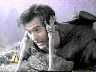 |
* The great mystery of how osteoblasts and osteoclasts cooperate so that there will be just the right amount of bone is on its way to being solved. CBFA1 is the factor that osteoprogenitor cells make to turn themselves into osteoblasts (Bone 25: 107-8, 1999); its levels decrease markedly with age. The endosteum (quiet bone-lining) cells make RANK-L, a membrane-bound protein factor that binds to RANK on osteoclast precursors and turns them into osteoclasts. Busy osteoblasts make osteoprotegerin, which interferes with the interaction by soaking up the RANK-L. Obviously something is missing from the picture; since bone grows along lines of stress, it may be an electrical phenomenon. See JAMA 292: 490, 2004; NEJM 353: 918, 2005. Update on osteoclasts: Am. J. Path. 170: 427, 2007.
Remember that interleukin-6 is a strong osteoclast activator; this probably accounts for the bone wasting seen especially near joints with rheumatoid arthritis.
All collagen is "woven", but here the term has a special meaning. When bone is being made rapidly (unborn child, healing injury, some diseases), collagen fibers tend to be laid down as crisscross WOVEN BONE, made rapidly and able to withstand stress equally well in all directions. When things are more sedate, the new bone is laid down as LAMELLAR BONE, with collagen fibers nearly parallel, which takes longer to make but is much stronger. They are easy to tell apart using polarized light; after age 3, woven bone indicates pathology.
The slim CEMENT LINES (blue on H&E) that you may see in bone have a little less collagen and a little more other stuff. They are found in remodeled bone at the border where new bone meets old, and are probably there to absorb impacts and make it harder to fracture the bone.
The osteoblasts are also responsible for beginning the process of mineralization, but this can't be the whole story, since a row of osteoblasts typically overlies an unmineralized "osteoid seam", which then takes about two weeks to be mineralized under the influence of the same cells.
Bone is decalcified prior to histologic sectioning, but bone that was calcified is less homogeneously dense-pink than bone that was never calcified ("osteoid"). Since it takes about two weeks to mineralize osteoid well, wide osteoid seams beneath the osteoblasts mean bone is being laid down fast or not being mineralized. Narrow osteoid seams mean the osteoid is being laid down slowly.
You can label the osteoid that mineralized today by administering tetracycline. This will impart a longstanding yellow color and distinct fluorescence to the seam. (Yes, if you were on tetracycline for a while for your acne, your skeleton was much more yellow and much more fluorescent than your friends'.)
|
Parts of the long bone (i.e., a bone that formed from a cartilage model):
Diaphysis... The long shaft Epiphysis... Between the growth plate and the nearest joint Metaphysis... Between the growth plate and the diaphysis. In kids, this is where most of the bone growth is taking place, so this is where most pediatric bone disease (infections, tumors) will occur. | 
|
When osteoblasts are working (growth, healing fractures, bone disease), they produce extra alkaline phosphatase, which shows up in the serum. This also forms the basis of the classic BONE SCAN, which lights up osteoblasts.
You can spot dead bone under the microscope because there are no osteocytes in the lacunes.
In vitamin C deficiency (scurvy), osteoid fails to form. In vitamin D deficiency (rickets / osteomalacia), osteoid fails to mineralize. (Future radiologists: In kids, look for sawtooth rib-cartilage junctions in both deficiencies.) Most Americans are vitamin D deficient by the classic standards; With today's fad diets, especially when forced on children (and they are, even some of the incredibly stupid ones), you are likely to run into serious diet-related bone disease.
The "brown tumors" of hyperparathyroidism feature areas of excessive osteoclast activity; these regress when the hyperparathyroidism is treated.
There's considerable bony variability among normal people.
Stature, of course, is entirely the result of bone growth (and, in age, of bone deterioration). Stature is mostly genetic; you know the effects of poor nutrition, chronic disease, hGH excess and deficiency, and osteogenesis imperfecta; precocious puberty will first make you taller than your peers, then your epiphyses close and you may end up shorter in the long run. It's actually the estrogens that close the epiphyses: Lancet 357: 1723, 2001, which is why we give an aromatase inhibitor (to prevent estrogen synthesis) when we treat boys with constitutional delayed puberty. If you get your sex hormones late (or never -- XXY's for example), you're likely to be tall. Rich people have been giving their sons growth hormone to make them taller for over two decades -- there has been no obvious harm.
A missing or extra (i.e., cervical) rib is generally a mere incidental finding. A missing phalanx or even radius isn't particularly disabling. Various anomalies of bone formation are a radiologist's and geneticist's fascination.
* Bone "cysts" are uncommon but do occur, usually as fluid-filled round cavities near active growth plates. They are lined by a simple layer of bone-lining cells; old reports that they were not lined resulted from these being missed after the lesions were curetted out.
![]() Epidermoid inclusion cyst
Epidermoid inclusion cyst
"Solitary bone cyst"
NJ Med Pathology -- Case study
{18649} polydactyly
FRACTURE TERMS
STRESS FRACTURE: The bone fractures after repeated extra stress, i.e., there were some tiny microfractures first and the bone was already a bit sore
INCOMPLETE FRACTURE: The bone is cracked, but not broken into two pieces. The best-known incomplete fracture is the fast-healing GREENSTICK FRACTURE from an impact to a child's supple long bone.
CLOSED (SIMPLE) FRACTURE: The overlying tissues are intact
OPEN (COMPLICATED) FRACTURE: The bacteria have a route from the surface to the bone; perhaps the bone is even sticking out the wound.
MULTIFRAGMENTED FRACTURE (formerly "comminuted fracture"): The bone is broken into several pieces.
COMPLEX FRACTURE: A curious term for a fracture in which the ends of the bone fragments have done serious damage to the surrounding tissue.
PATHOLOGIC FRACTURE: Due to intrinsic disease of the bone; the force would not have broken a normal bone. Seen in osteoporosis, cancer, osteogenesis imperfecta, others.
PROCALLUS: Fibrin / granulation tissue bump forming from the hematoma at the fracture site. "Provisional callus".
CALLUS: Granulation tissue bump at the fracture site that is starting to turn into fibrous tissue and cartilage, from which healed bone will arise. Eventually becomes BONY CALLUS and then remodels to look more or less like the original bone.
PSEUDARTHROSIS: The ends of the bone did not heal back together with bone. At best, there is fibrous scar connecting the ends, and a "false joint" is created.
If the fracture is multifragmented, if the ends of the bones are much displaced, or if infection happens, don't expect good healing. Likewise, poorly-nourished people or osteoporotic people are likely to get bad results.
![]() Clavicular pseudarthrosis (trust me)
Clavicular pseudarthrosis (trust me)
Prize photograph
Institute of Medical Illustrators
* Future forensic pathologists: Don't be fooled by post-mortem "heat fractures" of bones in burned bodies. These are caused by boiling of the bone marrow.
{07029} healing fracture, histology
{08994} healing fracture, histology
|
|
|
Four days: Periosteal new bone formation beginning
Ten days: Fracture line no longer visible; soft callus present
Fourteen days: Hard callus present
Twenty-one days: Callus is all hard
Ninety days: Bone has remodelled
* Orthopedic nails and screws placed into bone become separated from the bone itself by organized connective tissue continuous with the periosteum.
* Allogenic stem cells to enhance bone healing: Impressive animal study, no immunosuppression required J. Bone. Surg. 85: 1927, 2003.
OSTEOGENESIS IMPERFECTA ("brittle bone diseases", * fragilitas ossium; Clin. Ortho. 401: 6, 2002; Lancet 363: 1377, 2004)
A family of diseases having in common defective synthesis of type I collagen.
* The molecular biology has been extensively worked out (Clin. Orth. 343: 23, 1997); often there are too many mutations replacing glycine with some other amino acid. Here are the classic subtypes:
type I... Make too little pro-α1(1). Normal statue, lax joints, hard of hearing (another mutation: J. Clin. Invest. 85: 282, 1990).
type II... Pro-α1(1) is too short, etc. The collagen is made but poorly secreted. Bones break in utero or during birth, killing the child (yet another mutation: J. Clin. Invest. 83: 574, 1989)
type III... Triple helix doesn't form well. Short, many fractures, progressive kyphosis, hard of hearing, bad cases are lethal in childhood
type IV... Pro-α2(2) is too short, etc. Short stature, somewhat fragile bones
And if you look hard, you find many "forme fruste" people whose bones are just a bit more fragile (J. Clin. Invest. 97: 1035, 1996).
Some other genetic diseases prodce a similar phenotype but are not at the COL1A1 or COL1A2 loci.
Most are autosomal dominant; type II's are either autosomal recessive or new autosomal dominant mutations. (Explain.) In most cases, the teeth are deformed.
Except for type IV, all feature distinctive blue sclerae. (The "blue" color results from the translucence of the collagen. Don't rely on this, and don't go calling every relatively dark sclera "blue").
Bisphosphonates (which suppress osteoclast function / induce osteoclast apoptosis) for osteogenesis imperfecta: After some early flops, finally success. NEJM 339: 947, 1998; Pediatrics 111: 573 & 601, 2003, Lancet 363: 1427, 2004, others; pamidronate seems to be standard now and obviously works (J. Ped. 148: 456, 2006; J. Ped. 149: 174, 2006; J. Clin. Endo. Metab. 91: 511 & 1268, 2006; Pediatrics 119(S2): S-163, 2007, lots more).
Bone marrow transplantation as a treatment for osteogenesis imperfecta: Nat Med. 5: 309, 1999. This did not become widespread. Gene therapy and somatic cell therapy are under investigation (Clin. Orth. 379-S: S126, 2000).
|
{12402} osteogenesis imperfecta
{15795} osteogenesis imperfecta, blue sclera {18255} osteogenesis imperfecta type II {15801} osteogenesis imperfecta type II {18256} osteogenesis imperfecta, x-ray {15813} osteogenesis imperfecta, sutures not present |  "Unbreakable" increased public awareness of osteogenesis imperfecta. Mr. Jackson is rather tall to be an OI patient. |
Achondrogenesis II: Lethal mutation in COL2A1
* Osteogenesis imperfecta must be ruled out in children with
fractures attributed to abuse. This usually isn't difficult;
today, the full panel of labs seems to be urine organic acids,
serum amino acids, vitamin D, parathyroid hormone,
ceruloplasin/copper for Wilson's (seems far-fetched),
and (to diagnose osteogenesis imperfecta), sequencing of the
COL1A1 and COL1A2 genes (Clin. Ped. 49: 78, 2010).
The riddle is what to do with a kid with a mild mutation and
a situation that still looks like inflicted injury.
I used to be asked about "temporary brittle bone disease"
and after examining the evidence, decided that I don't believe in it.
Shortly afterwards, the physician who "discovered" this went
to court for making stuff up (Br. Med. J. 328: 187, 2004).
OSTEOPETROSIS ("marble bones", "Albers-Schonberg disease";
Am. Fam. Phy. 57: 1293, 1998)
This family of genetic diseases features progressive obliteration of the marrow cavity by bone
because the osteoclasts don't work. In severe cases,
(1) the bones become very brittle (think -- it's easier to crack something solid
than something porous); (2) the patient is likely to die of neutropenia or
anemia; (3) the bone impinges on the cranial nerves.
Most of these diseases are autosomal dominant (usually fairly mild)
or autosomal recessive (usually severe).
.
Other autosomal recessive patients lack carbonic anhydrase II, and
the osteoclasts die of too much acid (Hum. Genet. 99: 634, 1997;
Blood 97: 1947, 2001).
The dominant form is caused by a faulty chloride channel, preventing
the cytoplasm from becoming sufficiently acidic to dissolve bone in the first place
(Am. J. Path. 164: 1537, 2004; J. Clin. Endo. Metab. 92:
771, 2007). Of course, the forme fruste is resistance to osteoporosis, and
this has now been demonstrated (J. Clin.
Endo. Metab. 91: 995, 2006).
In the severe, autosomal-recessive variants, the skull becomes so deformed from the disease itself and
the extramedullary hematopoiesis that the foramina for the optic and other cranial nerves are
compromised. Radiologists note a striking resemblance between skull x-rays of these kids and the
"Alien" from the Sigourney Weaver space thrillers (Radiology 183: 129, 1992).
There are several autosomal dominant osteopetrosis genes; as you'd expect,
most feature excessive numbers of non-functioning osteoclasts, while a deficiency in
functioning RANKL produces osteopetrosis without osteoclasts (Nat. Gen. 39: 960, 2007).
As you would expect, the problem seems to be with the osteoclasts. If you get a chance to examine
these, they are likely to look bizarre.
Marrow transplantation cures the underlying lesion: Blood 97: 1947, 2001;
its use is now widespread for severe variants Ped. Clin. N.A. 57: 171, 2010.
"Osteopetrosis tarda" is a forme fruste, usually a subclinical x-ray finding
in older folks (Am. Fam. Phys. 57: 1293, 1998).
An infectious cause has been hypothesized but not found.
The achondroplasia locus is the receptor for fibroblast growth factor 3.
* The mutation is usually the same, a substitution: Nat. Gen. 13: 233, 1996. Other, worse alleles here give
thanatophoric dwarfism, with excessively short ribs and suffocation shortly after birth (update Am. J. Path. 161: 1325, 2002).
Rat model
Nature Genetics 12: 390, 1996. Some short people have a forme fruste ("hypochondroplasia")
with mutated FGF3/FGFR3 (J. Ped. 133: 5, 1998). Sun Hudson,
the baby in the 2005 Texas
"life support" case, suffered from thanatophoric dwarfism.
Also at the FGFR3 locus are some genes for craniosynostosis, in which the
sutures fuse too readily.
Advanced paternal age is one of the known risk factors for new mutations.
Apparently this mutation, and maybe the similar Apert's mutation (see below)
give a growth advantage to the sperm clone (Science 301: 606 & 643, 2003).
{25610} achondroplasia
* Several of history's best-loved entertainers were achondroplastic dwarves (legendary gladiators, the
Egyptian good-luck spirit Bes, Pharaoh Pepi's Nubian dancer, Tom Thumb and his wife, the great
Renaissance fiction-writer Morgante. The "Munchkins" from the movie
version of "The Wizard of Oz" were apparently
a mix of pituitary and achondroplastic
dwarves. Some people
say that Aesop was a black
achondroplastic dwarf -- you can chase down the classical sources yourself.
The traditional teaching is that
dachshund dogs are achondroplastics; we now know that the FGFR3 genes seem
to be normal, and the gene(s) for the short legs of dachshunds, basset
hounds, and bulldogs remains unknown (Can. J. Vet. Res. 64: 243, 2000).
* In fact, achondroplasia has a mystique, and has even been considered a desirable trait. See Clin.
Genet. 37: 279, 1990. Everyone's heard of the "Little People"'s organizations, and there is much
friendly rivalry between achondroplastic and pituitary dwarves as to "which is better", etc.,
etc.
There are several other dwarfism syndromes that result from defective bone growth. Those that
compromise the lengthening of the ribs are fatal shortly after birth (why?)
* Apert's (deformed face, syndactyly; the official name is
"acrocephalosyndactyly") is mutated fibroblast growth factor receptor 2 (Nat. Genet. 13:
48, 1996); documentary "Mary Ann" about an Apert's baby who was so ugly that everybody
assumed she was retarded, too, and they put her in a home for retarded kids; she wasn't retarded.
Apert's is usually a new mutation, and almost always on Dad's chromosome (Nat. Genet. 13: 9,
1996); this is another of the few genetic diseases that gets more common with advanced paternal age.
A milder allele causes Crouzon syndrome; the same advanced paternal age effect
has been noted (Am. J. Hum. Genet. 66: 768, 2000).
One of the "pseudoachondroplasia" diseases
results from a mutated cartilage matrix protein,
which is improperly processed and causes apoptosis of the growth cartilage
(Am. J. Path. 163:
101, 2003).
* Historically, there was an attempt to call normally-proportioned
very-short people "midgets" and people short because of disproportion
"dwarves". The word "midget", promoted by P.T. Barnum, is now "considered offensive" because of the
way language evolved.
{53757} Apert's
Here is the evidence: All this fits with pycnodysostosis and probably nothing else.
Making the call: JAMA 191: 111, 1965; more recently Nat. Genet. 10: 128, 1995.
This is an autosomal recessive illness caused (at least sometimes)
by defective cathepsin K, found only in osteoclasts and responsible
for removing bone matrix (J. Clin. Endo. Metab. 85:
425, 2000); the bones are demineralized normally by osteoclasts but they
cannot take out the collagen fibers. It's not clear exactly how the phenotype is produced.
Pathology J. Clin. Endo. Metab. 89: 1538, 2004.
* OTHER GENETIC SYNDROMES
Camurati-Engelmann disease, or "progressive diaphyseal dysplasia",
features bone laid down under the periosteum; the gene is TGF-β1
(Nat. Genet. 26: 273, 2000;
J. Biol. Chem. 278: 7718, 2003).
BONE INFARCTS
Infarction and necrosis in the shaft is more difficult because of collateral
circulation, and usually results from trauma. Eventually, a bone infarct becomes radiodense. This is probably
because of dystrophic calcification of the dead marrow-fat.
Remember osteonecrosis of the femoral head
("femur head necrosis", formerly "avascular necrosis" or "aseptic necrosis")
(J. Bone Joint Surg. 88A: 1117, 2006). It's a dread complication
of sickle cell disease, decompression sickness, and of course femoral neck fracture or
(as in Bo Jackson's case) dislocation. There is a mysterious link
to alcohol abuse, lupus, HIV infection
and glucocorticoid use (* apoptosis of osteocytes is the cause
at least sometimes -- Rheumatology 52: 235, 2013)
Anabolic steroids, once reputed to be a risk factor, seem to have been
exonerated.
* King Tutankhamun's necrotic metatarsal heads: JAMA 303: 638, 2010.
PYOGENIC OSTEOMYELITIS (Lancet 364: 369, 2004)
Common bacteria can reach the bone via the blood, during surgical or other trauma, or from
surrounding tissues (don't forget infected teeth, or the gangrenous feet of diabetics).
Osteomyelitis is especially serious, since the rise in pressure caused by the suppuration (like in a ripe
pimple, of course) is often enough to cause infarction of spongy bone and marrow, sequestering
the infection and turning it into a chronic infection refractory to all but the most aggressive
treatment.
The most common bug is staph The anatomic pathology of pyogenic osteomyelitis has many variants. Rupture through the cortex
via Haversian and Volkmann canals, will
produce PERIOSTITIS or (in a growing child) a SUBPERIOSTEAL ABSCESS surrounding the entire shaft.
Dead cortical bone becomes a SEQUESTRUM (and acts like any other foreign body; consider surgical
debridement), while new living
bone that grows around the dead bone
is called the INVOLUCRUM. Rupture through the skin produces refractory
pus-draining SINUSES, where
squamous skin cancer is likely to arise because of the ongoing destruction-and-regeneration
of the squamous epithelium. A walled-off area full of bacteria is a BRODIE'S ABSCESS,
which can keep the infection going, and perhaps spreading via the bloodstream, for many years.
In the chronic infection, the infiltrate is a mix of neutrophils,
lymphocytes, plasma cells, and lipid-laden macrophages; sometimes the plasma cells or
macrophages are by far the most abundant.
Look for numerous resorption pits in the dead bone or injured, signs of osteoclastic activity
at the end.
Healed osteomyelitis is often very radio-dense.
Remember that osteomyelitis is likely to complicate bedsores and foot gangrene.
Osteomyelitis often becomes a clinician's and patient's nightmare.
Amyloidosis A is one of the many feared outcomes.
* When there is an open wound that is being managed, pathologists are now being
asked to examine a bone biopsy to see whether osteomyelitis is present. This guides
antibiotic therapy (Am. J. Med. Sci. 321: 367, 2001).
Autopsy series of osteomyelitis in sacral decubiti: Arch. Path. Lab. Med. 127: 1599, 2003.
{05293} osteomyelitis, x-ray (see it? areas of dead bone often end up mottled-radiodense)
* SAPHO SYNDROME (synovitis, acne, pustules on the palmes / soles, hyperostosis, osteitis;
now, "acquired hyperostosis syndrome")
is a rare (and/or underdiagnosed?) illness in which
abscesses appear unpredictably in the skeleton. Patients also have acne.
We may reasonably think this is some kink in the immune system that prevents the body
from dealing with the propionobaterium that causes acne.
Bisphosphonates and antibiotics directed against the microbe are the mainstays of therapy.
Anti-TNF-alpha infliximab makes the synovial lesions much better and the
skin lesions worse: Rheumatology 45: 730, 2006.
SYPHILIS was famous for producing gummas and periostitis, and Shakespeare even calls it "the Neapolitan bone-ache."
TUBERCULOUS This used to be common everywhere and is still common in the poor nations. While the onset is
more insidious than pyogenic osteomyelitis, the infection is extremely destructive and hard to treat.
POTT'S DISEASE is the dread tuberculosis of the spine, and TB is the common cause of the infamous
PSOAS ABSCESS.
OSTEOPOROSIS (Ann. Int. Med. 126: 458, 1997; Med. Clin. N.A 87: 1039, 2003;
J. Clin. Inv. 115: 3318, 2005; Lancet 377: 1276, 2011;
molecules and cells J. Clin. Endo. Metab. 96: 600, 2011; J. Clin. Path. 64: 1042, 2011)
This is a very important process results from a slight excess of bone resorption over bone deposition,
continuing over many years. As we get older, we all get some osteoporosis.
The histology is banal (thin cortex, thin trabeculae), and the radiology equally so (there are
parameters on hand x-ray, and so forth). These belie the devastating consequences, which include
pathologic fractures (hip, compression fractures of vertebrae causing "dowager's hump" kyphosis),
and chronic pain. Osteoporosis causes 1.2 million fractures per year in the U.S. alone.
In a man, or a pre-menopausal woman, look for one of the known "causes" of "secondary
osteoporosis" (Arch. Int. Med. 149: 1069, 1989). These include, but are not limited to:
The first discovered gene was
the vitamin D3 receptor (* VDR gene product).
Original work: Nature 367: 576, 1994; Lancet 345: 423, 1995.
Update Ann. Int. Med. 145: 255, 2006 (some alleles probably do, others probably don't).
Also well-established is the presence of certain alleles at locus
for the estrogen receptor alpha. This interacts with VDR type (J. Clin. Endo.
Metab. 88: 3777, 2003).
Another gene turns out to be the one for type I collagen, where
certain alleles cause less bone mass and increase the risk for
osteoporosis (NEJM 338: 1016, 1998); collagen mutations can also promote
fractures independent of bone mass (i.e., the bone is poorly-made).
Several other less-well-known genes also seem important
(for example, a couple of lipoprotein-receptor-related genes Endocrinology 148: 2622, 2007;
JAMA 299: 1277, 2008).
Genetics updates:
J. Clin. Endo. Metab. 87: 2460, 2002; Lancet 371: 1505, 2008;
NEJM 358: 2355, 2008 -- includes osteoprotegrin, RANKL, RANK (no
doubt anymore J. Clin. Endo. Metab. 95: 3940, 2010), and the
estrogen receptor alpha -- not really surprising).
WNT1 -- NEJM 368: 1809, 2013; Nat. Med. 19: 179, 2013.
AB Plastin 3 (PLS3), a fairly severe X-linked syndrome with carriers at double the risk
of fractures: NEJM 369: 1529, 2013.
Although in "pure" osteoporosis, labs will be normal, it's probably worth
spending $75 to get each of these people a serum TSH (if on thyroid replacement), serum calcium,
serum PTH, serum 25(0H)vitamin D, CBC, chem profile,
and 24 hour urine calcium (J. Clin. Endo. Metab. 87: J. Clin. Endo. Metab. 87:
4431, 2002. This is now strongly supported, with a high percentage
of people with "their first fracture from osteoporosis"
also having vitamin D deficiency and/or low calcium
intake (extremely common), hyperthyroidism, and hypogonadism in men.
X-rays will be normal until about half of the bone is gone.
Today, "proven" ways of slowing osteoporosis include estrogen replacement (after menopause,
whether natural or artificial, see Am. Fam. Phys. 40: 205, 1989) and calcitonin (J. Ped. 118: 703,
1991). Androgens are the rule for men; bisphosphonates (osteoclast-inhibitors) became in the 1990's (South. Med. J. 87:
S-23, 1994).
Teriparatide, a recombinant form of parathyroid hormone, is now in widespread
use after its introduction in 2002.
Strontium, which slows osteoclasts and causes differentiation of matrix cells
to osteoblasts, seems to work well (Drugs & Aging 27: 711, 2010; J. Clin. Endo. Metab.
92: 3076, 2007).
Denosumab ("Prolia", one injection every six months), a monoclonal antibody against RANK-L (NEJM 354: 821, 2006), seems to work
at least as well as bisphosphonates, though it'll be pricey, and
will probably prove synergistic with
the bisphosphonates. Update Clin. Pharm. Ther. 91: 123, 2012.
Romosozumab is a monoclonal antibody that binds
to sclerostin, an osteocyte-produced inhibitorof
osteoblasts. It seems to be helpful in osteoporosis
(NEJM Jan 1, 2014).
*
Watch this one closely, as it will let us know about the future
direction of health care. If prophylactic denosumab therapy becomes commonplace,
the incidence of fractures in the elderly will drop dramatically.
In turn, this will cut dramatically into health care incomes.
* Odancatib inhibits osteoclast protease cathepsin K
(Toulouse Lautrec's protein).
There are now antibodies against two other proteins that inhibit
bone formation (sclerostin and the oddly-named dickkopf-1).
However, the idea that taking extra calcium even when you're a kid
is important because
it keeps you from getting osteoporosis years later is still dogma
(despite the meta-analysis Br. Med. J. 333: 775, 2006, which
reached the same conclusion based on clinical studies as I did by reasoning
from the basic pathology).
This government health scare remains politically correct, and
disclaimers are required.
("Recent research has raised
doubts about the efficacy of calcium supplementation in preventing fractures;
however, adequate calcium intake remains important." -- Am. J. Clin. Nutr. 85:
1361, 2007). Tell your patients "Uncle Sam still stays you need a lot
of calcium", just to cover yourself when they break a bone.
* Paradoxically, the amino-terminal end of parathyroid hormone
increases the formation and total mass of bone, and this is now
finding clinical use: NEJM 344: 434, 2001.
{46507} osteoporosis, gross
* There are other molecular mechanisms no doubt going on in osteoporosis.
In one mouse model, inadequate VEGF causes mesenchymal cells in bone to become
adipocytes rather than osteoblasts: J. Clin. Inv. 122: 3101, 2012.
Egyptian mummy ladies had much less osteoporosis for their ages than our ladies do; perhaps they were
more active physically than American folks today (Lancet 341: 673, 1993, classic Egyptian
art suggests that athletics was important in the culture and that healthy folks
weren't sedentary).
OSTEOMALACIA
Failure of the bone to mineralize properly in an adult.
Think first of inadequate intake of vitamin D and/or calcium.
This is a topic under "nutritional disease" and of course in childhood
it produces rickets.
Adults with dietary calcium deficiency (poverty, elderly "tea and toast" eaters,
people subsisting mostly
on vegetables) or malabsorption (especially, remember celiac sprue and post-bypass surgery
for obesity -- South. Med. J. 103: 570, 2010) are prone to osteomalacia, which in turn results in bone pain and
even fractures. In fact, about 90% of your patients with "osteoporosis-induced
fractures" turn out also to be vitamin D deficient and/or to have a low
calcium intake (J. Clin. Endo. Metab. 96: 1360, 2011).
And the truth is that adult vitamin D deficiency is rampant in our "civilized" society,
and accounts for many of your patients with persistent, nonspecific musculoskeletal pain
(Mayo Clin. Proc. 78: 1463, 2003). The skeleton will even light up on bone scan
(why?), and patients may be diagnosed with "metastastic cancer" -- this is
becoming common (Am. J. Med. Sci. 337: 245, 2009). Even people "who are not at risk"
turn up with it, and curiously, the "complementary medicine community" vitamin buffs
are paying this very little attention. Before your write your "total body pain"
patient off as "having fibromyalgia" or "being mental", check vitamin D levels.
* A study out of New Zealand (J. Am. Diet. Assoc. 104: 250, 2004)
followed 50 children who had avoided drinking cow's milk for a long time
and did not use calcium-rich supplements (i.e., the vegan or faddist
parents didn't know what they were doing).
They had almost three times as many fractures as other kids, most often
following trivial injury. This does not surprise me at all.
* There are some genes you don't have to learn yet.
HYPOPHOSPHATASIA, an inborn error that causes osteomalacia of
variable severity, is caused by mutations (recessive or a dominant poison-protein)
of alkaline phosphatase (J. Clin. Endo. Metab. 85: 743, 2000).
Of course, a mutated FGF23 causing overproduction of
its protein
causes osteomalacia ("autosomal dominant hypophosphatemic rickets").
Another osteomalacia gene that's probably fairly common is mutant
NHERF1, which governs resorption of phosphate by the kidney ("renal phosphate leak"; NEJM 359: 1128, 2008).
* Osteomalacia can also be caused by
paraneoplastic renal phosphate wasting:
NEJM 348: 1656 & 1705, 2003; JAMA 294: 1260, 2005;
"oncogenic osteomalacia" or "tumor-induced osteomalacia", from tumor-produced
FGF23 (fibroblast growth factor 23)
which you'll recognize
by the bone pain and the remarkably low serum phosphate levels; patients have sarcomas that
may be small, low-grade lesions
most notably the infamous "phosphaturic mesenchymal tumor": Arch. Path. Lab. Med. 126:
1245, 2002; JAMA 294: 1260, 2005; Am. J. Med. Sci. 332:142, 2006;
South. Med. J. 104: 348, 2011.
If you are looking at a section of bone that was made without the bone
being decalcified (which is hard to do), you'll see that the osteoid seams aren't well-mineralized.
More likely, you'll be looking at decalcified bone, and recognize
osteomalacia as very wide osteoid seams (the osteoblasts don't stop laying down
the collagen until they sense they've mineralized it).
It's a component of renal osteodystrophy; sorting this out is a mess we'll discuss under "kidney". OSTEITIS FIBROSA CYSTICA and RENAL OSTEODYSTROPHY
We'll cover these under hyperparathyroidism in "Endocrine", and when
we talk about bone disease in "Renal".
Remember that osteoclasts in
normal adult bone
are rare -- if you see even one osteoclast
in a random slide of adult bone, think of hyperparathyroidism from some cause.
Look for the cutting cones going through the centers of the trabeculae of spongy bone.
Renal osteodystrophy includes lesions of hypovitaminosis D, osteomalacia,
and hyperparathyroidism.
* FLUOROSIS
Nerve compression and increased radio-opacity are typical. You may see the
disease if you visit certain parts of Ethiopia or India (J. Bone. Joint Surg. 86: 594, 2004). Everybody in a particular
town will have it.
DIFFUSE IDIOPATHIC SKELETAL HYPEROSTOSIS is a common, usually asymptomatic process,
in which ligaments and tendons ossify, especially around the lower vertebral
bodies, linking them. They look like the drippings of wax candles.
PAGET'S OSTEITIS DEFORMANS (Am. Fam. Phys. 65: 2069, 2002; J. Clin.
Inv. 115: 200, 2005; Lancet 372: 155, 2008; NEJM 368: 644, 2013)
A common, usually-subclinical process seen in maybe 3% of older people, in which portions of one
or more bones become involved in abnormally rapid production and destruction of osteoid, leading
to curious, abnormally-vascular, abnormally-brittle bone that tends to deform along lines of stress.
Germline (variable expressivity) and sporadic mutations are now being sorted out (Endocrinology 152:
4180, 2011; J. Clin. Path. 63: 199, 2010); there are some syndromes at known loci
(Neurology 73: 636, 2009).
Most often involved are the pelvis (usually quiet), femurs (bowing of the legs), humerus (usually
quiet), spine (be careful) and/or skull. The forehead can grow larger ("leontiasis ossea").
The bone thickens and will feel warm because of
hyperperfusion. Patients may experience bowing of the legs, bone pain (usually mild) and increased
hat size. Deafness can result from impingement on the VIII nerve's foramina and/or disease of the
ossicles. The bones may fracture "like a chalk stick". Even more ominous are the (uncommon) development of high-output heart failure
(pagetic bone is very vascular and arteries communicate directly with veins)
and/or a
vicious osteosarcoma (or other bone cancer; fortunately only about 1% of patients develop
this; Cancer 70: 2802, 1992; Clin. Orth. 438: 97, 2005) and/or compression of the brain at the foramen magnum.
The microscopic picture is distinctive. Osteoblasts and osteoclasts
are both increased in number. Osteoclasts may be gigantic, with
100 or more nuclei.
Early, there are greatly increased osteoblasts and huge osteoclasts, with the marrow cavity
replaced by very vascular fibrous tissue. (This can cause high-output heart failure).
As the disease progresses, osteoid seam and woven bone get to be more prominent.
Late, the trabeculae are thick, made of woven bone, and shaped
weirdly, with a mosaic pattern of seams / cement lines ("geographic bone"; "crazy quilt". late in the disease)
where osteoblasts have filled irregularly-shaped, giant resorption pits.
A famous, ghoulish but helpful
autopsy-table observation: the
calvarium of a Paget's skull doesn't hold water.
Putting together what we know about the etiology of Paget's: J. Clin. Inv. 115:
200, 2005. The fundamental lesion is that osteoclast precursors are too sensitive
to factors that transform them into osteoclasts. At the beginning, the process
is entirely lytic, but soon the osteoblasts, which remain coupled, catch up.
Nobody knows why Paget's tends to be focal.
The idea that Paget's is a slow-virus infection -- once very popular --
seens to be discredited.
* The most impressive positive studies
(for example, a supposed trademark strain of measles --
J. Bone Min. Res. 17: 145, 2002) remain unconfirmed,
most genetic studies have turned up negative, and a
review team found contamination in one major lab reporting
positive results (J. Bone Min. Res. 22(S1: S-281, 2007).
It is likely that Beethoven's deafness was due to Paget's. (Why do we think so? Hint: Note the
shape of his head, and read up on his later-life health problems.)
* Egil the Viking, who acquired a hideously deformed, massive face and head, and whose skull
survived a blow from an axe from another Viking (who knew how to use axes),
probably had Paget's, which was common (Sci.
Am. 272(1): 82, 1995). Great pictures.
There are several familial syndromes, and sporadic cases feater
milder mutations in these alleles (J. Clin. Path. 63: 199, 2010).
The two genes worth knowing (SQSTM1 and TNFRSF11B/osteoprotegrin)
both interact with RANK and NFkappaB. Egil's severe familial
disease was probably SQSTM1. See NEJM 347:
175 & 210, 2002; Arth. Rheum. 50: 1650, 2004; many more.
The treatment of Paget's has been revolutionized by the introduction of the bisphosphonates,
osteoclast inhibitors (Br. Med. J. 312: 454, 1996; Hosp. Pract. 32(3): 63, March 15, 1997).
Don't confuse "Paget's disease of bone" with "Paget's disease of the nipple" or "Paget's disease of the
skin". The latter two result from growth of underlying adenocarcinomas into the epidermis.
{13384} Paget's disease, skull, gross
HYPERTROPHIC OSTEOARTHROPATHY
Mysterious periosteal new bone formation at the distal ends of tubular bones
throughout the body, with arthritis.
* Usually these patients also have clubbing, which
probably reflects megakaryocyte embolization in most cases. However,
something else is perhaps going on in hypertrophic osteoarthropathy.
Most of these patients have an underlying
non-oat-cell
bronchogenic carcinoma or cystic fibrosis. (Other notable causes of
clubbing, most especially right-to-left cardiac shunts, SBE, and Crohn's disease,
usually don't
cause the hypertrophic osteoarthropathy). Some cases are "primary", idiopathic,
progressing over decades.
This can be very painful and crippling, and require special treatment
(drugs, radiation). * Pamidronate in cystic fibrosis: Chest 121: 1363, 2002.
Historically, it was seen in radium painters and workers with white phosphorus ("phossy jaw").
It has always been known as a complication of osteopetrosis.
It has returned as a known risk to patients treated with high doses of bisphosphonates (Clin. Tox. 45: 753, 2007),
especially if they are also on glucocorticoids (J. Oral. Max. Surg. 68:
1055, 2010).
The common denominator seems to be whatever slows down osteoclasts.
It's puzzling why the upper and lower jaws are selectively involved.
I suspect that the team that identified bacterial biofilms is on the right track,
and that the cause will be found to be biofilm formation around dead bone that is not
removed on time by osteoclasts.
(J. Oral Max. Surg. 66: 767, 2008).
* Bones are a perennial symbol of human mortality, and in a larger sense, of all of human biology.
"A soft tongue can break hard bones": Proverbs 28:19.
"Bone of my bone..." -- Adam. "Cursed be he who moves my bones" -- Shakespeare's epitaph.
"Them bones, them bones gonna rise..." -- Afro-American Spiritual. As a kid, I was haunted
by Yeats's cryptic ghost tale The
Dreaming of the Bones. As an Irishman, Yeats couldn't actually say, "Isn't
it time we forgave the English?", but the very-short piece
is recommended reading in any era of political hatred.
INTRODUCTION TO BONE AND SOFT TISSUE TUMORS
Primary tumors of bone by definition arise from the mesenchymal cells (as opposed to the marrow
elements, plasma cells, and so forth). Soft tissue tumors by definition arise from
mesenchyme not part of bone or blood. If malignant, primary bone and soft tissue tumors
are called sarcomas. Benign or malignant, they are especially troublesome.
The GOOD news about many of these hated tumors is the triumph of high-intensity focused
ultrasound ablation as a means of destroying tumors in order to save limbs.
(This is also the "big news" in treating prostate cancer.) Review Radiology 255: 967, 2010.
In fact, amputations for sarcomas are uncommon nowadays and usually happen when it's been
neglected and the limb is already ruined: Bone & Joint Surg. 95B: 127, 2013.
The common primary bone cancers (as opposed to metastatic and hematopoietic cancers including myeloma) are osteosarcoma
(most common), Ewing's sarcoma, chondrosarcoma, and malignant giant cell tumor.
Among all cancers, only 0.2% or so arise in bone, with an incidence of 1/100,000.
The World Health Organization's nomenclature is standard.
Sarcomas are famously common as second cancers within old radiation ports.
Radiation-induced sarcomas are more aggressive than non-radiation-induced sarcomas
with the same histopathology, but are far from hopeless (Cancer 118: 2682, 2012).
When we are discussing bone tumors and mention "osteoid", we are referring
to a lace-like pattern of collagen deposited in the tumor, like spongy bone.
Where do bone tumors arise?
Diaphysis: enchondromas; some chondroSARComas; Ewing's, and eosinophilic granulomas
Epiphysis: chondroBLASTomas (famous trivia item); many giant cell tumors (supposedly)
Metaphysis: all other primary bone tumors (why? because this is where a tumor
arising from the growth plate will appear)
Osteomas arise from the cortical bone of the face. Plasma cell myeloma produces its "punched-out"
lesions throughout bone.
Most patients with primary bone cancer are young. While any primary bone tumor can occur in a
child or adolescent, remember these general ranges:
Metastatic neuroblastoma: infants and toddlers
Ewing's sarcoma: older children and adolescents
Osteosarcoma: adolescents and young adults
Giant cell tumors: young adults and middle age
Chondrosarcoma: middle age
Metastatic cancer: middle and old age
Cancers present with pain/tenderness, swelling, and/or a fracture. Benign tumors, if symptomatic at all, usually
present as a painless mass. (Osteoid osteomas are painful, enchondromas may cause a stress
fracture, and other benign tumors can sometimes do these things.)
The more aggressive cancers look like other cancers, and there is usually destruction of surrounding
bone.
Radiologists suspect cancer whenever a tumor lifts up the periosteum. You can tell because this
results in new bone formation (Codman's triangle; "sunburst" deeper in the bone).
Risk factors for bone sarcoma include some familial syndromes, previous radiation, and
previous chemotherapy
Cancer 67: 193, 1991). There is soft evidence of a risk for malignant fibrous histiocytoma
of the knee after knee infarct (Clin. Ortho. 467: 1820, 2009). However, most cases occur without any of these.
Unlike carcinomas, there's more known about chromosomal breaks than point mutations in these tumors.
(I suspect this is because the mutagens are more likely to reach epithelial cell nuclei than reach connective tissue / bone
cell nuclei.)
In the previously irradiated patient, the commonest primary bone cancers:
Tall kids get more Ewing's sarcomas and osteosarcomas.
Most bone tumors are slightly more common in males than in females. Chondrosarcomas in
particular are a man's disease (3:1).
Making the diagnosis of a primary bone tumor poses special problems.
The pathologist will always want to see the x-rays before making the diagnosis. (Lytic or blastic?
What sort of edge? Reaction in surrounding bone?)
Most cases will go for consultation. Few community pathologists have much experience with these
things.
There are a bunch of non-neoplastic foolers (Arch. Path. Lab. Med. 136: 772, 2012).
The tumors mostly look very similar anyway. It is easy to miss the tumor on biopsy. The
patients are kids. The treatment is horrible. What if she is pregnant?
* Biopsying these tumors: Clin. Orthp. Rel. Res. 368:
212, 1999. Open biopsy is the gold standard;
Tru-Cut core needle biopsy is now most common.
Fine needle aspiration of bone: Clin. Orthop. Rel. Res. 373:
80, 2000; Cancer 90: 47, 2000, Am. J. Clin. Path.
111: 632, 1999; Am. J. Clin. Path. 115: 59, 2001;
Arch. Path. Lab. Med. 128: 759, 2004.
White knuckles.
Treatment for bone tumors is much better than in the old days.
Benign tumors may be treated by curettage and packing with bone chips from elsewhere.
Malignant tumors require resection, radiation, and/or chemotherapy.
* Targeted therapies that have been so helpful in many breast, colon, and lung
cancers are now starting to be used in incurable sarcomas. For example, in
sarcomas other than liposarcoma, the tyrosine-kinase inhibitor pazopanib may buy around two additional months
of life (Lancet 379: 1879, 2012).
* OSTEOPOIKILOSIS is a non-disease in which there are patches of extra-dense bone around
the skeleton. It fools the inexperienced into thinking there are osteoblastic metastases.
FIBROUS DYSPLASIA (Arch. Path. Lab. Med. 137: 134, 2013)
Since mutations are involved, this is best considered among the bone tumors.
In this condition, a portion of bone when the spongy trabeculae should be is replaced by fibrous
tissue in which poorly-formed spicules of woven bone are abundant. Simple fibrous dysplasia can
be MONOSTOTIC or POLYOSTOTIC.
Monostotic fibrous dysplasia is usually asymptomatic, a radiologist's curiosity.
Often one side of the jaw is involved, producing a distinctive asymmetry of the lower face.
Involvement of the shoulders and hips can produce disability. (* Ask a radiologist to show you the
"shepherd's crook" deformity of the proximal femur.)
* Leave the variant "osteofibrous dysplasia" of the lower leg bones to us; it can be more debilitating (i.e., bow legs) but often self-cures.
MCCUNE-ALBRIGHT POLYOSTOTIC FIBROUS DYSPLASIA is a curious disease also featuring café-au-lait spots
(irregular borders, in contrast to those of neurofibromatosis), precocious puberty, and often other
endocrine dysfunctions (notably hyperthyroidism, cushingism, acromegaly, and/or vitamin D
resistance). It looks like a genetic disease, but isn't inherited in any familiar fashion.
The defect is in the gene that codes for the 5α-subunit (GNAS-1) of the G-protein (i.e., the one
that operates from the cyclic-GMP/ras system) that is in charge of stimulating adenyl cyclase. This
means that when a cell is given a signal via cyclic-GMP, it responds as if it had been stimulated by
cyclic-AMP. This probably accounts for the endocrinopathies, but what about the spots?
It turns out these patients are all mosaics for affected and normal cells, and that only the places
where the cells bear the mutation are affected. (Even one dose of the McCune-Albright must be
lethal to the fertilized egg; the mutation is post-zygotic and clones of cells bearing the mutation are
distributed segmentally throughout the body and must go back to the not-very-many-cells stage.
Ask an embryologist.)
* How it was worked out: NEJM 325: 1688, 1991. There's also a
number of alleles at the same locus ("Albright's hereditary osteodystrophy") that can be inherited;
this should be familiar from "pseudohypoparathyroidism" and "pseudo-pseudohypoparathyroidism": J.
Clin. End. Metab. 76: 1560, 1993.
Regardless of etiology, the common problem in fibrous dysplasia seems to be activation of adenylate
cyclase, leading to c-fos overexpression, etc., etc. NEJM 332: 1546, 1995.
Transformation of fibrous dysplasia to cancer is rare.
While we're on the subject of scrambled bone growth: An
ANEURYSMAL BONE CYST is a
rapidly-expanding lesion with wide blood vessels. There may or may not be bone and/or giant cells
forming in the septa between the blood vesels.
These seem to expand a bone just like an aneurysm expands a blood vessel ("blowout expansion");
curiously, these can also "spread" across a joint. These can grow rapidly and
be very destructive.
Sometimes an "aneurysmal bone cyst" can be the first sign of some other
underlying tumor. And chromosomal abnormalities have been reported in even "ordinary" ones.
BONE-FORMING TUMORS
* An "endostosis" is a lump of cortical bone in the medullary cavity. It's a
common, harmless incidental finding on x-ray.
OSTEOMA: a lump of ordinary, dense/compact bone jutting off a head bone
(especially jaw or paranasal sinuses). A non-tumor; if multiple,
think of Gardner's.
Although they have no malignant potential,
they may impinge on the brain, obstruct sinus drainage, or just look ugly.
If multiple, suspect Gardner's disease (intestinal polyps, soft tissue sarcomas,
desmoids, epidermoid cysts).
OSTEOID OSTEOMA: a common benign tumor of osteoblasts.
A nidus of miniature bony trabeculae and fibrous tissue,
surrounded by very dense bone.
Occurs in the vertebrae or long bones of young adults, where it causes well-localized pain and
tenderness. The pain responds dramatically to aspirin (i.e., it must be
mediated by the very large amounts of prostaglandin E2 in these
lesions; Clin. Ortho. Rel. Res. 393: 258, 2001),
and the rim is also very heavily innervated
(Mod. Path. 11: 175, 1998).
Enucleation is curative, though of course difficult for physician
and patient alike. The patient will tell you
in the recovery room whether you got it all out.
The modern initial treatment is computer-guided thermocoagulation, with or without
a prior tissue diagnosis, by the radiologists (Radiology 224: 92, 2002). {10830} osteoid osteoma, histology
OSTEOBLASTOMA ("giant osteoid osteoma"; Arch. Path. Lab. Med. 134: 1460, 2010): a rare non-metastasizing but
locally destructive tumor of osteoblasts.
This usually arises in the vertebral bodies of young adults.
The tumor cells make new osteoid. All osteoblastomas are by definition larger
than 1.5 cm.
Cells in the center can look very active (though not terribly
anaplastic). However, they appear to "mature" at periphery.
This lets you know it's not an osteosarcoma, and likely to be better-behaved.
We won't ask you to make the distinction.
OSTEOSARCOMA: the commonest primary cancer of bone (not counting
plasma cell myeloma of course). There are about 6000 new cases in the US each year.
Cancer of the osteoblasts.
Pathology update: Am. J. Clin. Path. 125: 555, 2006 (great photos).
By definition, any and all tumors in which malignant cells
themselves directly
make osteoid are osteosarcomas.
Warning: Benign endochondral bone formation can take place in any cartilaginous tumor.
You must see the malignant cells themselves making the bone.
Warning: When the malignant cells are absorbed into the new bone
and become "osteocytes", they may look very benign.
Warning:
Remember also that normal bone surrounding any diseased area may show proliferative changes.
* The old term "osteogenic sarcoma" is ambiguous
and should be discarded.
* There are elaborate subclassifications. The old one was based on how much cartilage,
fibrous matrix, and recognizable osteoid the tumor was producing, and turned out to
have no bearing whatever on prognosis or treatment.
Don't worry about these.
Most osteosarcomas arise in the medullary
areas of the metaphysis of long bones (especially the knee; occasionally in the jaw or elsewhere; sometimes on
bone surface, sometimes
not even in bone: "extra-osseous osteosarcoma" Cancer 65: 2762, 1990). As noted, most patients are adolescents or young adults, and there is a slight male preponderance.
(Of course, many teenaged males have a painful knee, hence the delays in making the diagnosis.)
People in retinoblastoma families are at much greater risk, and deletions of the Rb anti-oncogene on
long arm of chromosome 13 are the rule in osteosarcomas. Li-Fraumeni (p53) families are also at
great risk.
"Secondary" osteosarcomas occur in old people with Paget's disease (especially in the pelvis), or
patients of any age with familial conditions with many osteochondromas and/or enchondromas. Paget's
of bone is very common in the elderly, and accounts for famous
"second peak" in osteosarcoma frequency during later life. Other "secondary" osteosarcomas follow radiation, chemotherapy, chronic osteomyelitis, bone
infarcts (caisson workers).
Osteosarcomas present variable histopathology.
Tumors are usually predominantly made of new bone, cartilage and fibrous tissue.
Less often, there may be a preponderance of vessels
("telangiectatic osteosarcoma", a variant easily mistaken
on conventional x-ray for aneurysmal bone cyst: Cancer 109: 1627, 2007;
Arch. Path. Lab. Med. 136: 572, 2012). * When there is
a preponderance of small cells in an apparent "osteosarcoma",
gene studies will usually be more typical of a Ewing's sarcoma.
Leave the diagnosis of "epithelioid osteosarcoma", "osteoblastoma-like
osteosarcoma", and "low grade central osteosarcoma" to us.
Grading of osteosarcomas has not proved very helpful for prognosticating
outcome, since almost all the "classic" ones
that arise in the medulla are anaplastic "high grade" tumors.
* For some reason (probably an initial mutation that's become widespread), it's
not uncommon for there to be multiple, simultaneous, high-grade primaries (Am. J. Clin. Path. 136:
799, 2011).
There are two important subtypes with a generally good prognosis and low grade that arise on
the bone surface.
JUXTACORTICAL ("parosteal") OSTEOSARCOMA: dense bone with a dense,
fibrous, only mildly-anaplastic stroma, usually at the distal femur.
Often it's hard to tell
you're even looking at tumor microscopically. There may be cartilage,
and the most treacherous
even carry a cartilage cap (i.e., pretending to be osteochondromas -- * tip from
the radiologists: a benign osteochondroma won't have a cartilage cap more than 2 cm thick Radiology 255: 857, 2010).
Juxtacortical osteosarcomas carry a good prognosis, unless there is de-differentiation (at presentation
or later) into a more aggressive sarcoma (Cancer 103: 2373, 2005).
PERIOSTEAL OSTEOSARCOMA, a rarity, is a ring of
clearly-malignant, calcifying cartilage-and-new-bone around a bone in a young
person. It may occur remote from bone: Arch. Path. Lab. Med. 115: 906,
1991. The prognosis is generally good and chemotherapy does not seem to be indicated (Eur. J. Cancer 41: 2806, 2005; update
Clin. Orth. 453: 314, 2007).
It's uncommon, but not unheard-of, for an aggressive osteosarcoma
to arise on the bone surface. These behave as do the common sort that
begin inside the bone (Cancer 85: 1044, 1999; update on "high-grade
surface osteosarcoma Cancer 112: 1592, 2008).
* Future pathologists: Immunostaining for osteoblast markers
(i.e., osteopontin, osteonectin, osteocalcin) isn't sufficient
to establish that a tumor is an osteosarcoma; for example, giant cell
tumor stroma often stains for these as well (Clin. Orth. 459: 8, 2007).
Once almost always fatal, the five-year
survival in osteosarcomas presenting
without metastatic disease and
receiving modern therapy is running
around 70%.
This has stayed stable for the past quarter-century (Cancer 115: 1531, 2009).
Today, the large majority of these young people, even with
high-grade tumors, keep their limbs (Clin. Ortho. 468: 2854, 2010).
It is quite acceptable to remove lung metastases surgically,
even several times (Cancer 104: 1721, 2005). These patients can still often
be cured this way.
{05755} osteosarcoma, x-ray
CHONDROMATOUS TUMORS (remember cartilage often undergoes dystrophic calcification)
OSTEOCHONDROMA ("exostosis", "ecchondroma"): "the commonest bone tumor", actually a hamartoma
A cap of normal cartilage on a bony stalk, usually near the growth plates. These grow up over time but usually stop when growth stops.
An incidental finding, with a slight potential for transformation into chondrosarcoma,
especially if multiple (Gardner's, or the familial exostosis syndromes).
However, any osteochondroma that continues to grow is suspicions for
transformation to chondrosarcoma (Clin. Orth. 411: 193, 2003).
* The gene EXT1 is known, and governs surface expression of heparan sulfate.
(Nat. Genet. 19:
158, 1998).
EXT2 has also been discovered
(Am. J. Hum. Genet. 62: 346, 1998). Both are probably
tumor suppressor genes. One sees these in the familial forms *for example, Cancer 82: 1657, 1998); usually they are NOT mutated in lone osteochondromas.
This is the bone tumor that is best-linked to previous trauma (i.e., a bit of a normal growth surface
got pushed over to the site where the tumor developed).
{05842} exostosis ("osteochondroma")
ENCHONDROMA (or just plain "chondroma"): a common oddity; a popcorn-shaped lump of cartilage inside the shaft of a bone
Most often involves the proximal phalanges. It is an incidental finding on bone scan or x-ray, or is
discovered when a child or athlete presents with a stress fracture.
Usually enchondromas are harmless.
If multiple (syndromes include
Ollier's generally with unilateral involvement, Maffucci's with
hemangiomas) or in large bones, there is about a 25% chance that
at least one will
transform into
chondrosarcoma -- and since these enchondromas may have a bit of atypia / a few multinucleated cells or whatever,
even when they are benign, you'll want sub-sub-specialist consultation.
Ollier's and Maffucci's must be anti-oncogene deletion syndromes but the
genes have
evaded discovery (Hum. Mut. 24: 466, 2004).
Ollier's is usually unilateral (i.e., post-zygotic mutation);
there is a distinct bilateral hands-and-feet-only variant (J. Ped. Ortho. 6: 15, 1997).
Malignant transformation: Hum. Path. 31: 1299, 2000.
* CHONDROMYXOID FIBROMA ("fibromyxoid chondroma"): a rare, benign spindle-cell tumor that
differentiates toward cartilage, with different regions
showing differing amounts of matrix that is almost hyaline cartilage, but not quite.
Occurs in the legs and feet of young adults.
This is a famous tricky-call
for the pathologist.
Future pathologists: Look for lobularity, with more cellularity at the edges than at the centers
Review,
emphsizing what's still not known: Clin. Orth. 439: 171, 2005.
* CHONDROBLASTOMA: a rare, benign tumor of primitive cells that stain as cartilage
Occurs mostly in the legs of young people.
Most are very cellular and tend to focal calcifications, and there may be a few mitoses.
A biphastic pattern (good cartilage, more primitive areas) may be seen.
The "chicken wire" pattern of calcification is famous.
You probably won't recognize cartilage, at least easily, but the tumor cells stain for S-100, the cartilage marker.
CHONDROSARCOMA: the second commonest bone tumor (again not counting
metastases or plasma cell myeloma); sarcoma of cartilage, with a true hyaline-cartilage matrix
Primary chondrosarcoma arises most often in the pelvis in middle-aged men.
Prognosis depends on the grade:
* Grade I: mild cellular atypia (slightly anaplastic
nuclei, binucleate cells, two cells in a lacune,
mitoses; especially, encasing a bone spicule on all sides helps distinguish it from a
frisky enchondroma, which is the tough call -- today, these tumors can be treated
with simple curettage and cryosurgery with a near-certain cure: Clin. Ortho. 468: 2765, 2010)
* Grade II: crowded cells, perhaps a few bizarre cells
* Grade III: nasty-looking (includes "mesenchymal chondrosarcoma", among the most treacherous
of sarcomas Arch. Path. Lab. Med. 136: 61, 2012; the only way
you may be able to make this hard call is by finding that it stains for type II collagen: Mod. Path. 18: 1088, 2005)
* Warning: Agreement among pathologists (even "bone tumor experts")
on the grade of a particular chondrosarcoma is notoriously bad: J. Bone Joint Surg. 89-A: 2113, 2007.
The Grade III lesions are by far the most likely to metastasize,
but when the lower-grade lesions recur locally,
the grade may be higher.
* For some reason, chondrosarcomas of the cricoid cartilage are fairly common.
Managing them is tricky.
* Low-grade chondrosarcomas seem to be treated adequately by curettage
and cryotherapy (freezing): Clin. Ortho. 468: 1956, 2010.
Chondrosarcomas are notoriously unresponsive to chemotherapy and even radiation. And the basic
molecular biology remains almost completely unknown. Update Orthop. Clin. N.A.
37: 9, 2006.
{05958} chondrosarcoma, gross
TUMORS OF UNCERTAIN ORIGIN
EWING'S SARCOMA: an extremely malignant tumor of uncertain histogenesis
but a well-studied genetic anomaly. Update J. Clin. Path. 56: 96, 2003;
pathologists seeking to distinguish it from its look-alikes see Arch. Path. Lab. Med. 131: 192, 2007.
Most patients are of European ancestry and in their teenaged years.
The tumor is painful, often presents as a fever and because the x-ray
also can loks like osteomyelitis, is often mistaken for this.
It arises in any bone, packs the nearby marrow without growing new bone,
disseminates widely and rapidly, and is the one bone
tumor that metastasizes readily to other bones.
It is composed of sheets of "small blue cells" with little cytoplasm (i.e.,
almost all nucleus, hence "blue"-staining), and the cytoplasm is usually loaded with
glycogen (unreliable for diagnosis, though). There is no stroma or reticulin between its large vessels, and the tumor is a viscous liquid,
like pus. There are mitoses, necrosis, etc.
Future radiologists: Instead of elevating the periosteum as "Codman's
triangle", the rapid growth through the cortex produces layers of calcification
("onion skinning").
* Future pathologists: Cells with pyknotic nuclei are interspersed among cells with live nuclei
in a Ewing's -- suggesting its rapid growth.
* A closely-related tumor, the "malignant peripheral neuroectodermal tumor",
has traditionally been distinguished from classic Ewing's sarcoma.
If it makes actual Homer-Wright rosettes (i.e., little attempts at neural
tubes filled with little nerve processes) and/or
makes at least two neural markers, it's one of these instead. For a comparison see Arch. Path. Lab.
Med. 118: 608, 1994. Since the distinction
seems not really to affect treatment or outcome,
today most pathologists do not distinguish
these entities, but call both Ewing's/PNET of bone or ("Ewing's Sarcoma Family",
including one other obscure entity). See below.
* You may hear the term "Askin tumor" applied to a similar tumor in the chest wall.
If caught early, the prognosis after radiation and chemotherapy is good, with 85% 5-year survival.
If it has spreads, chances for a cure drop substantially to about 25%.
Survival statistics are now similar for adults (Arch. Surg. 138:
281, 2003). Histopathology does not affect prognosis (i.e., there is no reason
to "grade" these tumors: Cancer 110: 275, 2007).
Today's pathologists help clinicians plan therapy by seeking the
gene that causes Ewing's (and its kin) in the peripheral tissues.
The chromosomal abnormality t(11;22); the usual fusion product antigen
is EWS/FLI1, and nowadays it is becoming the norm to "define"
"the Ewing's sarcoma family of tumors" as having a fusion gene
product involving EWS and a gene from the ETS family (which includes FLI1).
There is now a procedure using fluorescent in-situ hybridization
for the diagnosis of Ewing's/PNET that works on formalin-fixed,
paraffin-embedded tissue (J. Clin. Path. 58: 1051, 2005).
Today it is routine to look for this in marrow, even if
the disease seems localized (Cancer 100: 1053, 2004), to
plan therapy.
* The EWS gene, on chromosome 22, is involved in each of the the
trademark translocations causing the following rare sarcomas:
clear-cell sarcoma (EWS-AFT1: reviewed Arch. Path. Lab. Med. 131: 152, 2007),
desmoplastic small
round cell tumors (EWS-WT1, peritoneum), myxoid chondrosarcoma (EWS-CHN or NOR-1),
and some myxoid liposarcomas (EWS-CHOP).
Again, most pathologists emphasize the similarities between
Ewing's and "primitive neurectodermal tumor" as seen
elsewhere in the body.
Both have the famous (11;22) translocation.
PNET will have neural markers (neuron-specific enolase,
chromogranin, synaptophysin, S-100, Homer-Wright rosettes)
that you'd expect in a neuroblastoma as well as CD99/013 that you'd expect in Ewing's.
Ewing's tends to be more anaplastic-looking, too. There's a tendency to
call Ewing's "PNET of bone".
Pathologists also help prognosticate these tumors (as we do osteosarcomas)
by defining how extensive necrosis is following preoperative chemotherapy.
{49496} Ewing's sarcoma, resection specimen
GIANT CELL TUMOR ("osteoclastoma"): benign (sometimes locally aggressive,
rarely metastasizing)
malignant, a common spindle-cell tumor packed with
non-neoplastic osteoclasts bearing many nuclei. Review Orthop. Clin. N.A.
37: 35, 2006.
These arise in the long bones, usually in young adults. For some reason, the tumor is much more common in the Far East than elsewhere.
The tumor is mesenchymally derived,
and the actual neoplastic cells are covered with abundant
RANKL, causing
local non-neoplastic cells to transform into very large osteoclasts (Am. J. Clin. Path.
117: 210, 2002). Since it is loaded with osteoclasts, these tumors are
entirely lytic, and you will probably see no new reactive bone formation.
Histology is not a reliable guide to the risk for local
recurrence ("malignancy"). Occasionally
these tumors metastasize to the lungs; often the metastases
can be removed surgcically for cure.
* Future pathologists: Telling these from the brown tumors of hyperparathyroidism
and the "giant cell reparative granuloma of bone"
is a challenge. Leave it to us. We may check for CSF1 gene abnormalities. Denosumab, the anti-RANK-ligand therapy, helps in
unresectable / metastatic disease.
"Malignant giant cell tumor of bone" may make any of three things (Cancer 97: 2520, 2003):
Also remember any tumor of bone may sometimes be loaded with giant cells.
NON-OSSIFYING FIBROMAS ("fibrous cortical defects"; "metaphyseal fibrous defects")
Little hamartomas in the long leg bones of children and teens.
They are made of fibroblasts, stroma, and
(often) lipid-laden macrophages. If you look, you'll find them in 1/3 of all normal kids. They rarely
cause problems, and they go away by themselves.
* A juvenile ossifying fibroma has trabeculae rimmed with osteoblasts. These may be more aggressive.
UNDIFFERENTIATED PLEOMORPHIC SARCOMA ("malignant fibrous histiocytoma") OF BONE (Cancer 79: 482, 1997)
is the other important bone tumor, around 1/10 as common
as osteosarcoma.
Leave the diagnosis of this many-faced tumor to the pathologists.
We're following the World Health Organization's recommendation
for the nomenclature -- they like
"undifferentiated
high-grade pleomorphic sarcoma".
{21123} malignant fibrous histiocytoma, trust me
CHORDOMA: a rare, locally destructive tumor that arises from the notochord
These arise in the sacral and spheno-occipital areas at any age.
Histology shows myxoid intercellular areas with "physaliphorous" (bubbly) cells.
Though they are only "low-grade" (some say "benign") lesions and seldom metastasize,
the prognosis is guarded just
because of the location. Today's ultra-modern surgery and radiotherapy
are producing likely cures in the majority of cases (J. Neurosurg. 113:
1059, 2010).
METASTASES TO BONE
By far the commonest "bone cancer" if you want to consider it to be such, presenting as pain or a fracture.
The tumors that most commonly metastasize to bone are carcinomas of the lung, breast, prostate,
kidney, and thyroid.
Most commonly the vertebral bodies are involved; the distal extremities are usually spared.
"Blastic" and "lytic" lesions are distinguished. (Any primary can produce either type; prostate
metastases are the most likely to be blastic, because for some reason they acquire
the machinery of osteoblasts Cancer 116: 1406, 2010.)
Repair of damaged bone by osteoblasts is responsible for the positive scan and elevated alkaline
phosphatase.
REMEMBER: Plasma cell myeloma, malignant lymphoma (Arch. Path. Lab. Med. 133: 1868, 2009), and
eosinophilic granuloma (a low-grade tumor of dendritic macrophages)
are other important causes of "bone tumors".
SOFT TISSUE TUMORS AND QUASI-TUMORS
A plethora of benign and malignant tumors arise from the connective tissue of the support structures
of the body. As usual, the cells normally present in a tissue determine the possible cells of origin of
tumors.
There are about new 8000 primary soft tissue sarcomas in the US
each year (Cancer 91: 794, 2001). This represents only about 1%
of adult malignancies.
Most of these "just happen", and the cause is seldom clear.
There are familial syndromes -- for example, rhabdomyosarcomas
notoriously occur in Li-Fraumeni, Gorlin's, and neurofibromatosis.
Sarcomas that present a monotonous appearance have, as a rule, at least one
trademark translocation (Am. J. Surg. Path. 26: 965, 2002; assays Lab. Invest. 81:
905, 2001).
In fact, several soft tissue sarcomas are
now pretty much defined in terms of their trademark genetic signatures;
for
some reason, these are fusion proteins (Am. J. Path. 163: 691, 2003;
update Arch. Path. Lab. Med. 130:
1199, 2006). These are not the ones that tend to follow radiation.
Sarcomas that are
very pleomorphic usually lack a genetic signature, but have instead
very elongated telomeres (Am. J. Path. 164: 1523, 2004).
For adult soft-tissue cancers, grade is now known to be the most important
determinant for prognosis (Arch. Path. Lab. Med. 130: 1448, 2006). Here is the French grading system, which you should
be able to use:
Histologic grade: As a rule, the more superficial the tumor in the body, the better the prognosis.
* Scoring systems for pediatric sarcomas are slightly different; don't worry about them.
Mitotic count is especially important. (Cancer 116: 2266, 2010 -- St. Jude's).
Surgery is often more limited for these tumors than for malignancies elsewhere (i.e., you can cut out
a lung clean and easy, but not half of somebody's back.) One mark of malignancy, then, is local
recurrence.
These tumors intrigue pathologists, and we love to subclassify them.
Your instructor thinks it is
inappropriate for you to spend too much time studying all the differences among these tumors.
Here's what's worth knowing about the common ones.
* GRANULAR CELL TUMORS (mostly benign)
Tumors of schwann cell origin (S100-positive, etc.), made of polyhedral, PAS-D-positive
phagolysosomes.
This tumor is notable for inducing hyperplasia in overlying stratified squamous epithelium. (This
"pseudoepitheliomatous hyperplasia" can look much more disturbing than the tumor itself.)
These were formerly misnamed "myoblastomas" for no reason.
BENIGN FIBROUS HISTIOCYTOMA
A tumor of cells usually showing a mix of features of fibroblasts (spindle cells, collagen production)
and macrophages (* positive for α1-PI, lysozyme, factor XIIIa, etc.)
The most familiar benign fibrous histiocytoma is the banal DERMATOFIBROMA, a hard knot in the
dermis with pigmentation of the overlying epidermis. More about this under "Skin".
MALIGNANT FIBROUS HISTIOCYTOMA ("MFH")
Formerly "the most common soft-tissue sarcoma", most "MFH's" have been
reclassified according to their genetic and immunostain markers.
* A few "MFH's" survive. The "storiform" pattern is cartwheel, like ovarian stroma.
The "myxoid" pattern simply generates lots of ground substance. The "inflammatory" pattern is
chemotactic for polys (betcha it makes interleukin 8). "Dermatofibrosarcoma protuberans" (Am. J. Surg. Path. 36:
1897, 2012) is a
cartwheel variant with little malignant potential and a trademark translocation; a pigmented dermatofibrosarcoma
protuberans is a "Bednar tumor". Etc., etc.
{21146} malignant fibrous histiocytoma, gross (trust me on this and the following MFH's)
CELLULITE (* "gynoid lipodystrophy"; update Plast. Rec. Surg. 118: 510, 2006)
The histopathology is thinning of the dermis (perhaps from pressure
of increasing adiposity) and especially thickening
of the fibrous bands of the subcutaneous panniculus where the
edges of the "cellulite" form dimples. ("Scar contracts.")
See Am. J. Dermpath 22:
34, 2000; Plast. Recon. Surg. 103: 1934, 1998.
All recent controlled studies of non-surgical cellulite remedies have failed
to show any effect.
LIPOMAS
Benign tumors of fat, the most frequent soft-tissue tumor. Typically, a thinly-encapsulated bump of
subcutaneous fat that doesn't grow or regress with the rest of the fat.
{05810} lipoma, gross
* Most lipomas, and their benign and malignant variants, have chromosomal abnormalities involving the HMGA1 or
HMGA2 genes. In one family, the genetic mutation in patients with multiple lipomas, however,
was reported to be in the mitochondria (J. Am. Acad. Derm. 44: 132, 2001).
MDM2 at 12q15 is involved in translocations in benign lipomas, amplified in atypical lipomas and
well-differentiated liposarcomas.
Otherwise, the gene in the autosomal-dominant form has remained elusive (Israel Med. Assoc. J. 4: 1121,
2002; Cutis 79:
227, 2007).
HIBERNOMA ("brown fat lipoma") is a tumor of brown (primitive, mitochondrion-rich, several lipid
droplets per cell) fat. The term "hibernoma" comes from the role of brown fat in generating heat to
wake bears from hibernation.
{46354} hibernoma, histology
In ADIPOSIS DOLOROSA ("Dercum's disease"), which may be an anti-oncogene deletion syndrome or
mitochondrial mutation syndrome, the
patient has hundreds of lipomas that may hurt.
Whatever the cause, it remains remarkably elusive.
William Osler called this one of the three worst diseases of his time (with subluxation of the
sacroiliac joint and milk leg). Today we're treating it with liposuction (Ann. Plast. Surg. 33: 664, 1994).
At least in adiposis dolorosa (and we may conjecture, in other lipomas), the fat isn't burned very well
for energy (Clin. Sci. 81: 793, 1991).
* ANGIOLIPOMAS, rich in vessels often containing microthrombi,
are often multiple and often painful. There's a familial syndrome; the presumed
anti-oncogene has not yet been identified (Arch. Path. 123: 946, 1999;
Derm Online 13: 3, 2007).
* Let us worry about "atypical lipomas" / "atypical lipomatous tumors"
(odd fibroblasts but maybe no lipoblasts -- they now seem to be
lumped in with well-differentiated liposarcomas when you look at the molecular
markers amplified MDM2 and CPM --- Mod. Path. 22: 1541, 2009), "pleomorphic lipoma with
alaring-looking-but-benign floret giant cells", and so forth.
* "Lipoma of the spermatic cord" is a chunk of non-neoplastic fat in a man's inguinal hernia sac.
* MYELOLIPOMA is a bone marrow choristoma in or near the adrenal glands (review and case Arch.
Path. Lab. Med. 118: 188, 1994).
{25412} adrenal myelolipoma
* ANGIOMYOLIPOMAS are the familiar tumors that, when present in the kidney, raise the specter of
tuberous sclerosis.
* LIPOBLASTOMAS are curious, lobulated tumors of children
that look like myxoid liposarcomas minus the mild anaplasia. They are
often multiple. Let us worry
about them. There is a trademark rearrangement at 8q12 of the PLAG1 gene; a lipoblastoma
will probably get sent for this to distinguish it from a myxoid liposarcoma.
Neither lipomas nor liposarcomas have any know relationship to obesity.
LIPOSARCOMA ("liposarc"): The most common cancer arising in adult soft tissues
"Cancer of the fat", though capable of arising wherever there are blood vessels (which give rise to fat
in the baby). They usually arise deep in the body, especially in the retroperitoneum.
* The classification of liposarcomas has undergone recent revision.
The pathology team at Columbia
correlates each of the four types with the genes activated
during a particular stage of maturation of fat (fascinating: Am. J. Path. 172: 1069, 2008).
"Well-differentiated liposarcomas" feature normal fat divided by septa containing
some atypical cells, possibly with lipoblasts. There are fewer than 5 mitotic figures
per ten high power fields. Prognosis depends on location. Retroperitoneum and groin are bad,
extremities is good.
"Myxoid liposarcomas" look grossly like raw fish fillets. They feature a plexiform pattern of
vessels in a myxoid background with little lipoblasts. * "Round cell liposarcomas"
used to be described; most are a mix of areas
that look initially like lymphomas, except that at least
a few cells contain fat
vacuoles, mixed in with some myxoid liposarcoma. Since they also feature
the trademark t(12:16) translocation, the WHO classification (2002)
recommends they be considered lumped with myxoid liposarcomas as "myxoid / round cell" for studies
(as is now done; Cancer 109: 2522, 2007).
However, the more round-cell liposarcoma
in a tumor, the
more aggressive. Most often, the round-cell area is found within a myxoid liposarcoma
and imparts a more ominous prognosis.
"Pleomorphic liposarcomas" blend into totally anaplastic sarcomas. The give-away is huge, ugly
cancer cells loaded with fat vacuoles. Update Cancer 117: 5359, 2011.
* There's also immunostain markers: MDM2 and CDK4 are well-established as
markers for malignancy in tumors of fat-cell origin
Am. J. Clin. Path. 125: 693, 2006; J. Clin. Path. 62: 1127, 2009.
* "De-differentiated liposarcomas" are less-well-differentiated non-lipoblast-containing sarcomas
that arise in well-differentiated liposarcomas (Cancer 106: 2725, 2006).
{05804} liposarcoma, gross
RHABDOMYOMA
Very, very rare. Most "rhabdomyomas" you'll see are the cardiac hamartomas typical of tuberous
sclerosis. Often the cells are rich in glycogen (they're muscle), with the nucleus
looking like the body of a spider in a web.
RHABDOMYOSARCOMA ("rhabdo": Arch. Path. Lab. Med. 130: 1454, 2006)
Cancer with skeletal-muscle differentiation (i.e., sarcomeres on electron microscopy, maybe cross-striations on light
microscopy, positive for desmin and myoglobin, etc.)
They are the most common soft tissue sarcomas of children; if localized,
cure rates are about 75%, but less than 20% if metastatic; a variety of
targeted therapies are in the works (J. Clin. Onc. 28:
1240, 2010). Less often,
rhabdomyosarcomas occur in adults (Cancer 91: 794, 2001). Surprisingly, only a minority of these cancers arise in the muscles (probably because skeletal muscle
is pretty much post-mitotic). They can arise most anyplace else (notably the head and neck or the
GU tract).
"Embryonal rhabdomyosarcoma" usually occurs under age 20.
Look for "tadpole cells" and "strap cells" with cross-striations, and "spider cells", with a nucleus
surrounded and distorted into a spider shape by vacuoles of glycogen.
* There is often allelic loss at 11p15; the gene is not yet known.
Prognosis is now quite good, with about 80% survival.
"Botryoid sarcoma", now removed
from the category of "embryonal rhabdos", has
a benign overlying epithelium layer and a packed
"cambium" layer beneath. This uncommon tumor occurs in the urogenital tracts of children.
The prognosis is better than for other rhabdomyosarcomas, with around 90% survival.
* "Spindle cell rhabdomyosarcoma" has also been removed from the
category of "embryonal rhabdos"; its prognosis is generall good.
"Alveolar rhabdomyosarcoma" is very aggressive. The cells have scanty cytoplasm, and
have very little septa; the term "alveolar" derives from the
presence in some of these tumors of a very fine vascular support
system resembling the alveoli of the lungs. This cancer most often strikes
teenagers. The gene mutation to remember is PAX. * Trademark translocations (1:13) or (2;13) bring either PAX7 or PAX3 adjacent to
FKHR/FOX01 (J. Clin. Onc. 20: 2672, 2002 -- PAX3 translocations are more common and more ominous)
Update J. Clin. Inv. 122: 403, 2012. "Pleomorphic rhabdomyosarcoma" is similar to, but is much more anaplastic than, an "embryonal
rhabdo". You won't see cross-striations, but you will probably see big,
deeply-eosinophilic cancer cells. It is less common than either embryonal or alveolar,
and patients are usually adults (in contrast with the other rhabdomyosarcomas). It is the most aggressive of all.
* Future pathologists: Myogenin and MyoD1 are two of several
popular
markers for rhabdo differentiation.
{24748} rhabdomyosarcoma (striations)
LEIOMYOMA
Tumors made of interlacing bands of benign smooth muscle. The bands present
the familiar whorled appearance on section.
You'll see plenty of leiomyomas in the uterus ("fibroids") and GI tract.
However, they occasionally
occur away from large hollow organs.
A leiomyoma in the soft tissues often has arisen from blood vessels. These leiomyomas tend to be
painful.
Leiomyomas can have a variety of curious cellular changes, including
big hyperchromatic nuclei, and still be benign. Leave us to worry about this.
* Future pathologists: Usually it's an easy call, but if
the location is unusual, you'll often
make the diagnosis of leiomyoma/leiomyosarcoma
using α-smooth muscle actin immunostain.
LEIOMYOSARCOMAS
Cancer of smooth muscle. They resemble leiomyomas, but
"something's not right" -- either a very abnormal location (deep-in-the-flesh
is usually leiomyosarcoma rather than leiomyoma), or mitotic figures (even one in
twenty high-power fields probably means cancer).
Again, demonstrating alpha-smooth-muscle actin by immunostain is helpful in confirming smooth muscle origin.
Immunosuppressed transplant patients tend to develop smooth muscle tumors.
Epstein-Barr virus The prognosis for soft-tissue leiomyosarcoma is still generally guarded: Cancer 109: 282, 2007.
* The inferior vena cava is a fairly common site.
The prognosis is still poor (Am. J. Surg. 201: e18, 2011; J. Am. College Surg. 210:
185, 2010).
* Leave the distinction between true leiomyosarcomas
and "sarcoma with myogenic differentiation" to us (Cancer 118: 5349, 2012).
{21122} leiomyosarcoma, gross, femur (trust me)
* FIBROMAS
Surprisingly, true fibromas (i.e., tumors derived from
common fibroblasts) are rare. "Neurofibromas" arise from schwann cells (again, S100-positive) while "gum fibromas"
are reactive hyperplasias. Papillomas of the skin and
"fibroadenomas" of the breast have abundant stroma that may or may not really be neoplastic.
And so forth.
The "solitary fibrous tumor" (benign or malignant) is a distinct entity
that usually appears in the pleura but can occur almost anywhere. It's one of the "zebras" in the
differential diagnosis of hypoglycemia, as it can make an insulin-like factor.
Ask a pathologist to show you the "staghorn vessels".
FIBROSARCOMAS
Cancer of fibroblasts. Uncommon, and generally fairly well-differentiated.
Most fibrosarcomas feature the "herringbone" pattern shown nicely in "Big Robbins".
* A variant ("congenital fibrosarcoma" -- present at birth or at least announcing
itself during the first two years of life) features
a distinctive chrosomal translocation and fusion product, ETV6-NTRK3,
which is now assayed routinely (Am. J. Surg. Path. 24: 937, 2000).
{05828} fibrosarcoma, gross
* ELASTOFIBROMA usually occurs under the scapula and is perhaps
a hyperplasia due to unusual movement; there's little solid stuff on
genetics. It was originally described
as an occupational problem of jack-hammer operators; however, today the majority of
patients
are female. The pathologist sees dense fibrosis with spectacular elastic fibers, bead-strings,
and lumps. See Clin. Orthop.
Rel. Res. 387: 127, 2001; Ann. Thorac. Surg. 91: 1622, 2011.
SYNOVIAL SARCOMAS (* "Robert Urich's tumor")
A relatively common tumor that usually arises in the extremities
away from synovium, but that
usually shows
both epithelial and spindle-cell differentiation ("it's both an adenocarcinoma and a sarcoma"), as you would expect (why?)
It is now defined to have a t(X;18) translocation, which is rarely seen in other sarcomas.
Whether or not it really has anything to do with synovium (you decide for yourself),
you should remember that it's most common in young adults (ages 20-40), and among
them is the most common soft tissue sarcoma.
* Marker fusion oncogene: SYT-SSX (NEJM 338: 153, 1998; Am. J. Clin. Path. 111:
528, 1999), resulting
from any of three trademark t(X;18) translocations (SYT-SSX1 -- worse prognosis, SYT-SSX2, SYT-SSX4) that now seem to define the tumor.
We can stain for the oncoprotein to make the call.
p16INK4a/CDKN2a is often deleted (Am. J. Clin. Path. 126: 866, 2006).
There's a fairly specific immunostain SYT (Mod. Path. 20: 522, 2007).
An even more specific immunostain is TLE1 (Am. J. Clin. Path. 135: 839, 2011).
Prognosis depends on grade (mitotic figures, necrosis).
{10622} synovial sarcoma
We've already mentioned the major members of the FIBROMATOSIS family (Dupuytren's, Peyronie's).
* "Fibromatosis colli" will take out a sternocleidomastoid muscle, while little "fibromas" may stud a
newborn's fingers. Riedel's struma probably belongs here as well.
TRITON TUMORS
Benign or malignant, these exhibit both Schwann-cell and skeletal-muscle differentiation(!) Named
for the salamander that regrows its limb-buds.
Malignant triton tumor, by our alumnus Dr. Stasik: Arch Path. Lab. Med. 130:
1878, 2006.
DESMOIDS are aggressively invasive but non-metastasizing growths of fibroblasts. The most familiar
variant arises in the abdominal muscles during or after pregnancy. * Future pathologists: Tough call. They tend to light up with beta-catenin. Look for myofibroblasts
lined up in parallel.
* Watch for pegylated interferon alfa-2b as treatment: J. Am. Acad. Derm. 59(2S1): 57, 2008.
* NODULAR FASCIITIS (South. Med. J. 87: 842, 1994;
mutations are rare Am. J. Clin. Path. 131: 701, 2009;
needle biopsy Am. J. Clin. Path. 132: 857, 2009)
A mysterious, self-limited hyperplasia simulating a fibrosarcoma, with infiltration of soft tissue, lots
of mitoses, but no atypia. Extravasated red cells and a hypocelular center with a hypercellular
periphery are keys. Don't try to diagnose this one yourself.
INTRAVASCULAR FASCIITIS is nodular fasciitis growing down
a vein. It ends up looking like a sausage.
PROLIFERATIVE FASCIITIS ("proliferative myositis" when in muscle)
looks like nodular fasciitis with lone big (but not anaplastic)
pink cells interspersed
(look like ganglion cells).
TRAUMATIC MYOSITIS OSSIFICANS
A jock's disease, which usually follows a large bruise to a muscle. The hematoma organizes into a
tough fibrous mass that seems to infiltrate the muscle (or fat or other soft tissue) and usually exhibits
bony metaplasia.
Don't misdiagnose this as a frightfully aggressive sarcoma. Ask a lawyer whether this turns
malignant or whether injury ever really causes cancer (if it does, it's very rare).
Traumatic myositis ossificans from fraternity hazing: Clin. Ortho. 466: 225, 2008.
* INFLAMMATORY MYOFIBROBLASTIC TUMOR: Leave this stuff to us.
Most are benign; there's a common but not invariable rearrangement
involving ALK (Arch. Path. Lab. Med. 136: 623, 2012).
* SMALL ROUND CELL DESMOPLASTIC TUMOR is yet another sarcoma
with its own trademark translocation t(11,22). In this case, it's EWS/WT1.
Arch. Path. Lab. Med. 126: 126, 2002.
* EXTRASKELETAL CHONDROSARCOMAS are typically myxoid, and bear translocations
involving the nuclear receptor TEC. The common one is
t(9:22), producing the EWS/NOR-1 fusion protein; several others exist
(Am. J. Path. 162: 781, 2003; Cancer Res. 60: 6832, 2000).
ALVEOLAR SOFT-PART SARCOMA (J. Clin. Path. 59: 1127, 2006):
Its trademark PAS-positive crystals has so far been found to contain
only a few components, all obscure
proteins (Am. J. Path. 160: 1197, 2002). Its trademark genetic
lesion is a translocation between chromosome 17 and the X-chromosome; Am. J. Path. 159: 179, 2001); hence the risk
is double for girls (Cancer 103: 1245, 2005).
* It is slow-growing, but often lethal. Response to chemotherapy is poor.
{09873} alveolar soft part sarcoma, histology
* In some areas of longstanding pressure, where a decubitus might
ordinarily form, these fibroblasts can form a mass (Mod. Path. 6:
69, 1993; update on pseudosarcomas Arch. Path. Lab. Med. 132:
579, 2008). This has the name "atypical decubital fibroplasia" or
"ischemic fasciitis". Leave the diagnosis to us.
* Future pathologists: In immunocompromised people, mycobacterial infection can produce
a fibroblastic proliferation that's easy to mistake for a
fibrosarcoma with little anaplasia, especially if you don't think
to do an acid-fast stain. This is the infamous "inflammatory pseudotumor of
mycobacterial infection".
* Even more mysterious inflammatory pseudotumors -- big yellow masses --
can pop up in various
places around the body, notably the lung or wherever there is
urothelium. Pathologists look for spindle cells, often with
mitotic figures but without florid anaplasia. There will be a mix
of inflammatory cells, mostly lymphocytes and plasma cells; when
the latter are abundant, we talk about "plasma cell granuloma".
{11372} temporal bone and pons, normal
BIBLIOGRAPHY / FURTHER READING
I urge anyone interested in learning more about
bone and soft tissue pathology
to consult these standard textbooks.
In my notes, the most helpful current
journal references are embedded in the text.
Students using these during lecture strongly prefer this.
And because the site is constantly being updated,
numbered endnotes would be unmanageable.
What's available online, and for whom, is always changing.
Most public libraries will be happy to help you get an article
that you need. Good luck on your own searches, and again,
if there is any way in which I can help you, please contact me at
scalpel_blade@yahoo.com.
No texting or chat messages, please. Ordinary e-mails are welcome.
Health and friendship!
![]() Osteogenesis imperfecta
Osteogenesis imperfecta
X-rays from Harvard
* Other alleles at collagen loci:
Stickler's syndrome: Gawky people with some degree of nerve deafness; COL2A1 (the
major type II collagen gene) or another
collagen locus: J. Med. Genet. 36: 353, 1999
The most common recessive gene is a defective TCIRG1, an ATPase proton pump that
enables acid secretion by osteoclasts (Am. J. Path. 162: 57, 2003) and in
the stomach. Without it, the ability to absorb calcium is impaired, and patients
develop "osteopetrorickets", the marrow cavity filling with poorly-mineralized
bone (Nat. Med. 15: 610 & 674, 2009).
ACHONDROPLASIA (Lancet 370: 162, 2007; Arch. Dis. Child. 97: 129, 2012 especially for the new surgical options)
An autosomal dominant condition (often a new mutation) in which there is impaired formation of the
long bones by the familiar endochondral process. The limb bones are short, with abnormally wide
ends. The patient has a head and trunk of normal size, and disproportionately short but well-muscled arms and legs. The face
usually has a large forehead, prominent supraorbital ridges, and deepset root of the nose
(looks like a very mild Apert's).
Sexuality is generally normal, and intelligence is distributed as for the general population, with
many well above-average (Am. J. Med. Genet. 41: 208, 1991).

Don Sebastian de Morra, by Velazquez
The great danger to life is deformity at the foramen magnum,
where minor trauma can dislocate the skull from the neck bones and
compress the brainstem. However, this is rare and most
achondroplastic patients are not at risk. Review
and recommendations for screening: J. Neurosurg. 107(S2): 152, 2007.
{25688} achondroplasia
{49474} "achondroplasia" (looks like a thanatophoric dwarf to me, failure of rib development)
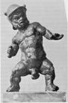
{53760} Apert's* PYCNODYSOSTOSIS
There seems to be a consensus today that artist Henri de Toulouse-Lautrec's
dwarfism and deformities were caused by PYCNODYSOSTOSIS.

Henri de Toulouse-Lautrec
There are a host of genetic syndromes involving bone, from
families where several members share a minor skeletal anomaly
to the horrible FIBRODYSPLASIA OSSIFICANS PROGRESSIVA (formerly "bad myositis
ossificans"),
probably caused by a mutated ACVR1, and lethal to the
fertilized egg (Nat. Genet 38: 35, 2006).
This causes overproduction of bone morphogenetic protein 4 in which wound
healing is accompanied by exuberant endochondral bone production.
![]() Fibrodysplasia ossificans progressiva
Fibrodysplasia ossificans progressiva
25 year old man
From NEJM
![]() Fibrodysplasia ossificans progressiva
Fibrodysplasia ossificans progressiva
Six year old girl
From Kaplan JBJSA 75A
Spongy bone can be infarcted easily wherever it has an end-artery pattern of vascularization
and the artery is compromised. This is most likely near the convex surfaces of joints.
The feared result is detachment of the articular cartilage (the dread "crescent sign" on x-ray).
The overlying cartilage and synovium / joint space will be spared.
![]() Osteonecrosis of the hip
Osteonecrosis of the hip
Bo Jackson's disease
WebPath Tutorial -- comments are down right now
![]() ,
though most of the common bacteria can produce osteomyelitis. In sicklers,
salmonella is the usual culprit; they are also prone to salmonella septic
arthritis (Clin. Ortho. Rel. Res. 468: 1676, 2010). Drug abusers who mix their works with tap water are prone to
pseudomonas. In 50% or so of cases, you'll grow nothing, probably because
somebody gave an antibiotic already.
,
though most of the common bacteria can produce osteomyelitis. In sicklers,
salmonella is the usual culprit; they are also prone to salmonella septic
arthritis (Clin. Ortho. Rel. Res. 468: 1676, 2010). Drug abusers who mix their works with tap water are prone to
pseudomonas. In 50% or so of cases, you'll grow nothing, probably because
somebody gave an antibiotic already.
{39505} osteomyelitis, x-ray
{40090} osteomyelitis, draining sinus
![]() Healing osteomyelitis
Healing osteomyelitis
WebPath Tutorial -- comments are down right now
* The other uncommon chronic focal osteomyelitis syndrome of children -- chronic
recurrent multifocal osteomyelitis -- is a mild disease in which children
seem to have osteomyelitis at various places but don't (Rheumatology 49: 1505, 2010).
It turns out there are no bacteria involved.
It may run with Crohn's regional enteritis,
and has responded to TNF-alpha blockers (Pediatrics 116: 1231, 2005).
Your lecturer is waiting for a genetic mutation
to be found, and suspects it's a lack of something that tells neutrophils that there's
already plenty in a bone (just as pyrin, which is lacking in familial mediterranean fever,
puts the brakes on neutrophil riots elsewhere.)
![]() Syphilitic periostitis, skull
Syphilitic periostitis, skull
Classic photo
Adami & McCrae, 1914
![]() OSTEOMYELITIS
OSTEOMYELITIS
* Literature buffs: Elizabeth Barrett Browning ("Sonnets from the Portuguese", etc.) was completely
disabled by severe chronic pain from (probably)
Pott's disease for over a decade. When she was given free
access to morphine, she became a productive writer and much happier person. Despite ideology,
there may be a lesson here.

Elizabeth Barrett Browning
![]() Pathology of Osteoporosis
Pathology of Osteoporosis
WebPath Tutorial
Heavy calcium intake (popular with the Tums manufacturers, of course) remains
"unproven" as a means of preventing the dread late effects of
osteoporosis. Children and teens are exhorted to take lots of calcium
to prevent osteoporosis in old age.
Not only are good current epidemiologic studies not there (meta-analysis BMJ 333: 775, 2006),
the idea also doesn't make sense -- osteoporosis reflects a lack of matrix protein,
not a lack of calcium.
Your lecturer believes that old work on plentiful calcium
preventing "bone loss" on x-ray resulted from its preventing osteomalacia (which
of course it often does), not osteoporosis. The two look the same on imaging studies. Go figure.
The genes that predispose to osteoporosis are being discovered.
{13844} osteoporosis, histology (thin trabeculae)
![]() Osteoporosis
Osteoporosis
WebPath Tutorial -- comments are down right now
![]() Osteoporotic compression fracture
Osteoporotic compression fracture
WebPath Tutorial -- comments are down right now
"Osteosclerosis" ("the opposite of osteoporosis") is not a specific illness, but a localized area in
which the trabeculae are too thick for whatever reason.
{46509} osteomalacia, thick unmineralized osteoid seams
{46510} osteomalacia, von Kossa stain for calcium (calcified is black, non-calcified is orange)
{12027} renal rickets
{12734} osteitis fibrosa cystica (osteoclast city!)
![]()
Certain locales have very high concentrations of fluoride ion in the
water. This is good for preventing dental caries (the teeth are very dark-mottled), but ultimately
causes hyperostosis of the skeleton.
The famous "paramyxovirus-like particles" seen in the osteoclast
nuclei have been spotted in a variety of other diseaes with abnormal
osteoclasts.
The same group that reviewed the viruses with negative results
also did not find somatic mutations of the known risk-genes in Pagetic bone.
Somatic mutations of SQSTM1 aren't present in sporadic Paget's
(J. Clin. Endo. Metab. 94: 691, 2009).
{18781} Paget's disease, skull
{09376} Paget's disease, bowed lower extremities
{09377} Paget's disease, bowed upper extremities
{38210} Paget's disease, histology
{18810} Paget's disease, histology
{13847} Paget's disease, histology
{13850} Paget's disease, polarized light; woven bone
![]() Paget's osteitis deformans
Paget's osteitis deformans
WebPath Tutorial -- comments are down right now
![]() Paget's osteitis deformans
Paget's osteitis deformans
WebPath Tutorial -- comments are down right now
* Worth mentioning here is another disease in which bone
mysteriously transforms, thankfully ultra-rare.
ESSENTIAL OSTEOLYSIS ("Gorham's disease") features replacement of bone (usually a single one,
or part of one) by cavernous hemangioma (or maybe just nothing: Virch. Arch. 442: 400, 2003). For some reason, these patients
also tend to get a chylothorax if the thoracic duct is involved.
Your lecturer suspects the cause is failure of osteoblasts to follow
the line of osteoclasts on the tail-end of the cutting cones.
OSTEONECROSIS OF THE JAW (Clin. Tox. 45: 753, 2007; Am. J. Med. 121: 475.e3, 2008; South. Med. J. 101: 160, 2008;
Ortho. Clin. N.A. 40: 223, 2009):
This is a once-and-future major disease.
The bone and/or marrow becomes necrotic.
The disease is focal, most often just behind the lingual side of the canine teeth.
 Sarcoma Images
University of Washington
Pictures and comments
Bone Tumors
NJ Med Path Dept
Nice tutorial
Bone Tumors
Henry DeGroot's site
X-rays and pathology
Here are two infamous trick pathology questions for rotations.
* One fooler in radiology is the "sports tumor" of young
people, elevated periosteum and tremendous osteoblastic activity on scan where the adductor muscles attach to the femur:
Am. J. Roent. 176: 1227, 2001.
Osteosarcomas and Ewing's sarcomas are typically
treated with chemotherapy before resection, and the more necrosis seen
in the resected specimen, the better the prognosis (robust finding Cancer 118: 5888, 2012).
![]() Fibrous dysplasia
Fibrous dysplasia
NJ Med Pathology
Case study
![]() Aneurysmal bone cyst
Aneurysmal bone cyst
WebPath Tutorial -- comments are down right now
![]() Aneurysmal bone cyst
Aneurysmal bone cyst
WebPath Tutorial -- comments are down right now
![]() Aneurysmal bone cyst
Aneurysmal bone cyst
WebPath Tutorial -- comments are down right now
* Results are widely variable. Some of the
failures probably were because the lesions weren't really
osteoid osteomas.
![]() Osteoid osteoma
Osteoid osteoma
WebPath Tutorial -- comments are down right now
![]() Osteoid osteoma
Osteoid osteoma
WebPath Tutorial -- comments are down right now
* ADAMANTINOMA of bone appears benign
clinically, radiographically, and histologically (it recalls tooth enamel cells),
but is locally aggressive and occasionally metastasizes. In the jaw, we call
these ameloblastomas. Otherwise, it'll be in the shaft of the tibia.
![]() Osteoid osteoma
Osteoid osteoma
NJ Med Pathology
Case study
![]() Osteoblastoma
Osteoblastoma
NJ Med Pathology
Case study
The tumor typically passes through the cortex, eventually eroding it, and
elevating the periosteum. "Codman's triangle" of new bone under elevated
periosteum is common.
* Rothmund-Thomson fragile-chromosome syndrome (RECQL4)
features an increased cancer risk, notably osteosarcomas (JNCI 95: 669, 2003).
Paget sarcomas are almost always high-grade and aggressive.
Around 90% are osteosarcomas, and 10% of them are non-osteosarcomas
(Arch. Path. Lab. Med. 131: 942, 2007).
{05906} osteosarcoma, gross
{05909} osteosarcoma, gross
{05927} osteosarcoma, gross
{05930} osteosarcoma, gross
{24747} osteosarcoma, gross
{21118} osteosarcoma, with pathologic fracture
{10322} parosteal osteosarcoma, gross
{05838} parosteal osteosarcoma, x-ray
{05864} osteosarcoma, histology
{25613} osteosarcoma, histology
{32123} osteosarcoma, histology
![]() Parosteal osteosarcoma
Parosteal osteosarcoma
WebPath Tutorial -- comments are down right now
![]() Clickable osteosarcoma
Clickable osteosarcoma
WebPath Tutorial -- comments are down right now
![]() Osteosarcoma
Osteosarcoma
WebPath Tutorial -- comments are down right now
![]() Osteosarcoma
Osteosarcoma
WebPath Tutorial -- comments are down right now
![]() Osteosarcoma
Osteosarcoma
Text and photomicrographs. Nice.
Human Pathology Digital Image Gallery
Cartilage Exhibit
Virtual Pathology Museum
University of Connecticut
![]() Osteochondroma
Osteochondroma
WebPath Tutorial -- comments are down right now
![]() Osteochondroma
Osteochondroma
WebPath Tutorial -- comments are down right now
![]() Osteochondroma
Osteochondroma
WebPath Tutorial -- comments are down right now
![]() Osteochondroma
Osteochondroma
WebPath Tutorial -- comments are down right now
![]() Enchondroma
Enchondroma
NJ Med Pathology
Case study
![]() Chondromyxoid fibroma
Chondromyxoid fibroma
NJ Med Pathology
Case study
![]() Low-grade chondrosarcoma
Low-grade chondrosarcoma
Soap bubble x-ray
NJ Med Pathology -- Case study
{09637} chondrosarcoma, gross
{46512} chondrosarcoma, gross
{49494} chondrosarcoma, gross
{09643} chondrosarcoma, lost its differentiation
{09649} chondrosarcoma, rib, gross
{10319} chondrosarcoma, pubis
{08997} chondrosarcoma, histology
{35999} chondrosarcoma, histology
{08998} chondrosarcoma, histology
![]() Chondrosarcoma
Chondrosarcoma
WebPath Tutorial -- comments are down right now
![]() Click on the chondrosarcoma
Click on the chondrosarcoma
WebPath Tutorial -- comments are down right now
![]() Chondrosarcoma
Chondrosarcoma
WebPath Tutorial -- comments are down right now
![]() Click on the chondrosarcoma
Click on the chondrosarcoma
CT scan
WebPath Tutorial -- comments are down right now
![]() Chondrosarcoma
Chondrosarcoma
WebPath Tutorial -- comments are down right now
* A final cartilage lesion worth learning (later) is
SYNOVIAL CHONDROMATOSIS ("synovial osteochondromatosis"),
in which metaplasia of synovium into balls of cartilage produces a picturesque,
mysterious lesion easy to diagnose on x-ray. Slightly atypical cartilage cells
may appear, especially near the edges.
* The search for an inhibitor of the gene product: Nat. Med. 15: 750, 2009.
{46408} Ewing's sarcoma, gross
{08470} Ewing's sarcoma, gross
{05812} Ewing's sarcoma
{38204} Ewing's sarcoma, histology
{40496} Ewing's sarcoma, histology (reticulin stain)
{46410} Ewing's sarcoma, small undifferentiated cells
{46411} Ewing's sarcoma, widespread necrosis
![]() Ewing's sarcoma
Ewing's sarcoma
WebPath Tutorial -- comments are down right now
![]() Ewing's sarcoma
Ewing's sarcoma
WebPath Tutorial -- comments are down right now
![]() Ewing's Sarcoma
Ewing's Sarcoma
NJ Med Pathology
Case study
![]() Giant cell tumor
Giant cell tumor
WebPath Tutorial -- comments are down right now
![]() Giant cell tumor
Giant cell tumor
x-rays
WebPath Tutorial -- comments are down right now
![]() Giant cell tumor
Giant cell tumor
WebPath Tutorial -- comments are down right now
![]() Non-ossifying fibroma
Non-ossifying fibroma
NJ Med Pathology
Case study
{21127} malignant fibrous histiocytoma, trust me ![]() Paget's disease
Paget's disease
Malignant fibrous histiocytoma
NJ Med Pathology case study
Pathology update: Cancer 98: 1934, 2003.
Molecular biology: Br. J. Cancer 98: 434, 2008. As a super-primitive tissue, they light with S-100
(unlike adenocarcioma) and
cytokeratin and epithelial membrane antigen (unlike a cartilage tumor).
![]() Osteoblastic metastases
Osteoblastic metastases
x-ray
WebPath Tutorial -- comments are down right now
![]() Osteoblastic metastases
Osteoblastic metastases
x-ray
WebPath Tutorial -- comments are down right now
![]() Osteolytic metastases
Osteolytic metastases
x-ray
WebPath Tutorial -- comments are down right now
![]() Osteolytic metastases
Osteolytic metastases
x-ray
WebPath Tutorial -- comments are down right now
![]() Bone metastases
Bone metastases
WebPath Tutorial -- comments are down right now
![]() Bone metastases
Bone metastases
WebPath Tutorial -- comments are down right now
![]() Eosinophilic granuloma
Eosinophilic granuloma
NJ Med Pathology
Case study
Soft Tissue Pathology
Sampurna Roy, MD
Lots of photos and good text
Lipomatous and Myxoid Tumors
Histopathology and essay
For pathologists
The best-known risk factor
is previous radiation (especially radiation therapy).
Extraosseous osteosarcomas occurring in an old radiation field
are all-too-familiar; also known from these sites are
malignant fibrous histiocytomas, then
are fibrosarcomas and angiosarcomas (Ann. Surg. 239: 903, 2004 -- radiation-induced angiosarcomas can be spotted by amplified myc).
Tumor differentiation:
Score 1: Looks almost like normal adult mesenchyme -- well-differentiated liposarcoma, well-differentiated fibrosarcoma (the ones without anaplasia), well-differentiated leiomyosarcoma (the ones without anaplasia)
Mitotic count:
Score 2: Familiar low-grade sarcoma (i.e., classic myxoid liposarcoma, common herringbone fibrosarcoma, myxofibrosarcoma, cartwheel MFH / dermatofibrosarcoma protuberans, common leiomyosarcoma, well-differentiated or common angiosarcoma)
Score 3: All the rest
Score 1: 0-9 mitoses per 10 high power fields
Tumor necrosis:
Score 2: 10-19 mitoses per 10 high power fields
Score 3: 20 or more mitoses per 10 high power fields
Score 0: No necrosis
Score 1: Less than 50% necrosis
Score 2: 50% or more necrosis
Grade 1: Total score 2,3
Grade 2: Total score 4,5
Grade 3: Total score 6,7,8* In contrast to osteosarcomas and Ewing sarcomas, the extent of necrosis
after chemotherapy given prior to resection does not have any implication
for prognosis: Clin. Ortho. 455: 219, 2007.
{09019} malignant fibrous histiocytoma, histology
{09020} malignant fibrous histiocytoma, histology
{09021} malignant fibrous histiocytoma, histology
{09119} malignant fibrous histiocytoma, electron micrograph; see the macrophage features?
![]() Malignant fibrous histiocytoma
Malignant fibrous histiocytoma
Tom Demark's Site
The familiar uneven skin dimpling on the buttocks and thighs. Women are
much
more likely to be affected, nobody knows why.
{12172} lipoma, gross
{12171} lipoma, gross
{15460} lipoma, colon
{40056} lipoma, gross
![]() Lipomas
Lipomas
Text and photomicrographs. Nice.
Human Pathology Digital Image Gallery
* The link with mutated mitochondrial
DNA was first noted in the 1990's: Am. J. Hum. Genet. 52: 551, 1993.
It is now well-established, and there are several known mitochondrial-DNA / lipoma
syndromes (Neurology 52: 1103, 1999).
HIV-ASSOCIATED LIPODYSTROPHY SYNDROME is a well-known problem faced by patients on long-term
retroviral therapy. No one really knows how it happens. It features redistribution of
bodyfat from the subcutaneous tissues into the skeletal muscles, development of
insulin resistance, and very low levels of adiponectin (J. Clin. Endo. Metab. 88:
627, 2003). The mainstay of therapy is intensive exercise, both aerobic and
resistance; metformin may be helpful. In addition to the curious way in which
exercise distributes existing fat back to normal locations, a man on antiretroviral
therapy who loses his bodyfat by diet-exercise looks like any other "ripped / defined" man.
{25413} adrenal myelolipoma
The prognosis for well-differentiated and myxoid liposarcomas is generally
good; for the others, poor. Surgery is the only modality that helps.
Distinctive for liposarcomas
is the presence
of at least a few lipoblasts. These are mesenchymal cells with hyperchromatic nuclei
with fat droplets
that scallop the nuclei (less often, one big fat droplet that greatly indents the nucleus).
You don't see these in healthy tissue, even in
embryonic or fetal fat, or in benign tumors of fat.

If you have a sharply-demarcated area in which another sarcoma (or maybe carcinoma) has arisen, you're into
"dedifferentiated liposarcoma", much more ominous.
* The usual translocation t(12;16; FUS-CHOP / DDIT3) fusion product is well-characterized (update Am. J. Clin. Path. 168: 1642, 2006;
less often FUS/TSL is uninvolved and EWS fuses to CHOP/DDIT3).
Now that the trademark translocation has been discovered, a variety of poorly-classified low-grade
sarcomas have been found to be myxoid liposarcoma variants (Am. J. Clin. Path. 137: 229, 2012).
{05807} liposarcoma, gross (a big one!)
{10607} liposarcoma, gross
{09004} liposarcoma, histology
{24750} liposarcoma, fat stain
![]() Rhabdomyoma
Rhabdomyoma
Pittsburgh Pathology Cases
All are thoroughly
malignant, and even with today's therapies many patients die.
{10955} rhabdomyosarcoma (straps and tadpoles)
{16703} rhabdomyosarcoma, spindle-cell variant
{16704} ditto, electron micrograph
{09006} alveolar rhabdomyosarcoma, histology
{24858} sarcoma botryoides, gross and x-ray
{25303} embryonal rhabdomyosarcoma
{25304} embryonal rhabdomyosarcoma
{25305} sarcoma botryoides, cambium layer
![]() Rhabdomyoblast
Rhabdomyoblast
Nuclei at edges
ERF/KCUMB
![]() Leiomyoma
Leiomyoma
Electron micrographs
VCU Pathology
![]() appears to be the cause (NEJM 332: 19, 1995; this gets
confirmed from time to time, for example Hum. Path. 33: 133, 2002).
appears to be the cause (NEJM 332: 19, 1995; this gets
confirmed from time to time, for example Hum. Path. 33: 133, 2002).
{05834} leiomyosarcoma, gross (trust me)
{25357} leiomyosarcoma, gross (trust me)
{09016} leiomyosarcoma, histology
{05831} fibrosarcoma, gross
{16680} synovial sarcoma, good biphasic histology
{40604} synovial sarcoma, biphasic
* The APC locus is likely to be mutated, and a few other common
chromosomal lesions are described. Not surprisingly, familial
adenomatous polyposis and Gardener's are risk factors for desmoids (Cancer 74: 1270, 1994).
In fact, when I began residency, our world-famous soft-tissue tumors expert said he would
never sign out nodular fasciitis without consultation with a second pathologist.
At the end of my residency, our chief told us a story of an alum who, as a junior attending
pathologist, self-diagnosed his own sarcoma and had his arm amputated, only to learn
later that it was just nodular fasciitis. You have been warned.
A rare young-person's sarcoma, worth mentioning
to medical students because of its curious biology.
* Pathologists use the TFE3 stain to spot the translocation.
{09875} alveolar soft part sarcoma, PAS stain showing crystals
{09876} alveolar soft part sarcoma, PAS stain showing crystals
![]() "Osteoarthritic" lipping
"Osteoarthritic" lipping
WebPath Tutorial -- comments are down right now
FRISKY FIBROBLASTS
Future pathologists need to be on guard against calling "sarcoma!" on
large fibroblasts with hyperchromatic nuclei that they'll sometimes
see in the craters of ulcers (gut, decubiti -- "atypical decubital dysplasia", etc.).
 * SLICE OF LIFE REVIEW
{11756} bone, normal
{11757} bone, normal
{11758} cartilage, normal
{11759} cartilage, normal
{12064} hand & wrist bones, severe joint erosion abnormal joint congruity
{12206} bone marrow, normal
{12263} bone marrow, normal
{12266} bone marrow, normal
{13619} bone marrow aspirate, normal
{14630} hyaline cartilage (lacunae)
{14631} hyaline cartilage (lacunae)
{14632} hyaline cartilage, normal
{14633} hyaline cartilage, normal
{14634} hyaline cartilage, normal
{14635} hyaline cartilage, normal
{14636} elastic cartilage, normal
{14637} elastic cartilage, normal
{14638} elastic cartilage, normal
{14639} cartilage, calcifying (electron micrograph)
{14640} cartilage, calcifying (electron micrograph)
{14641} fibrocartilage, normal
{14642} fibrocartilage, normal
{14643} fibrocartilage (polarized), normal
{14644} osteoblasts (developing bone), normal
{14645} osteoblasts (developing bone), normal
{14646} osteoclasts (howship's lacunae), howship's lacunae
{14647} osteoclasts (howship's lacunae), howship's lacunae
{14648} osteoblasts (active), normal
{14649} osteoblasts (active), normal
{14650} bone, cortical
{14651} bone, cortical
{14652} bone, adult cortical
{14653} bone, adult cortical
{14654} haversian system in formation
{14655} haversian system in formation
{14656} resorption cavity, bone
{14657} resorption cavity, bone
{14658} osteon (forming)
{14659} osteon (forming)
{14660} cortical spongy bone, normal
{14661} cortical spongy bone, normal
{14662} cortical & spongy bone (polarized), cross section with polarized light
{14663} cortical & spongy bone (polarized), cross section with polarized light
{14664} osteoblast ultrastruct., electron micrograph
{14665} osteoblast ultrastruct., electron micrograph
{14666} osteoclast, normal
{14666} osteoclast, normal
{14667} osteoclast, normal
{14668} osteoclast ruffled border (electron micrograph)
{14669} osteocyte (electron micrograph), normal
{14670} intramembranous bone formation, normal
{14671} intramembranous bone formation, normal
{14672} osteoclast ruffled border (electron micrograph)
{14673} osteoclast ruffled border (electron micrograph)
{14674} primary spongy bone formation
{14675} primary spongy bone formation, normal
{14676} bone, development of typical long bone
{14677} bone, development of typical long bone
{14678} tetracycline label of bone-forming site
{14679} tetracycline label of bone-forming site
{14680} spongy bone, modeling
{14681} spongy bone, modeling
{14682} trabecular bone resorption
{14683} trabecular bone resorption
{14684} bone lining cells
{14685} bone lining cells
{15121} cartilage, hyaline
{15123} cartilage, elastic
{15124} cartilage, elastic
{15126} cartilage, fibrous
{15130} cartilage, fibrous
{15131} cartilage, fibrous
{15132} cartilage, fibrous
{15133} bone, puppy
{15134} bone, puppy
{15135} bone, puppy
{15136} bone, puppy
{15137} osteoblast in puppy bone, #8
{15138} bone, * cartilaginous
{15139} bone, * cartilaginous
{15140} bone, * cartilaginous
{15141} cartilage, articular
{15142} cartilage, articular
{15143} bone, cement
{15144} bone, cement
{15301} bronchus, cartilage
{15918} bone growth plate, normal
{16343} temporal bone, normal
{20285} bone, normal
{20737} cartilage, hyaline
{20738} cartilage, hyaline
{20739} cartilage, elastic
{20740} cartilage, elastic
{20741} cartilage, elastic
{20742} cartilage, elastic
{20743} cartilage, fibrous
{20744} cartilage, fibrous
{20745} bone, osteocyte
{20746} bone, osteoclast
{20747} bone, intramembranous bone formation
{20748} bone, endochondral bone formation
{20749} bone, endochondral bone formation
{20750} bone, endochondral bone formation
{20751} bone, endochondral bone formation
{20752} bone, endochondral bone formation
{20753} bone, endochondral bone formation
{20754} bone, endochondral bone formation
{20755} bone, endochondral bone formation
{20901} cartilage, elastic
{20908} cartilage, hyaline
{46393} osteogenesis imperfecta, normal
{48426} skull, normal
Enzinger and Weiss's Soft Tissue Tumors
Kilpatrick's Diagnostic Musculoskeletal Surgical Pathology
Robbins and Cotran Pathologic Basis of Disease
Rosai and Ackerman's Surgical Pathology
Rubin's Pathology: Clinicopathologic Foundations of Medicine
Demay's Cytopathology
Sternberg's Diagnostic Surgical Pathology
Wold's Atlas of Orthopedic Pathology


| New visitors to www.pathguy.com reset Jan. 30, 2005: |
Ed says, "This world would be a sorry place if
people like me who call ourselves Christians
didn't try to act as good as
other
good people
."
Prayer Request
Teaching Pathology
 Drop by
and meet Ed
Drop by
and meet Ed
If you have a
Second Life
account, please visit my teammates and me at the
Medical Examiner's office.
PathMax -- Shawn E. Cowper MD's
pathology education links
Ed's Autopsy Page
Notes for Good Lecturers
Small Group Teaching
Socratic
Teaching
Preventing "F"'s
Classroom Control
"I Hate Histology!"
Ed's Physiology Challenge
Pathology Identification
Keys ("Kansas City Field Guide to Pathology")
Ed's Basic Science
Trivia Quiz -- have a chuckle!
Rudolf
Virchow on Pathology Education -- humor
Curriculum Position Paper -- humor
The Pathology Blues
 Ed's Pathology Review for USMLE I
![]()
![]()
 | Pathological Chess |
 |
Taser Video 83.4 MB 7:26 min |
 |
Click here to
see the author prove you can have fun skydiving without being world-class. Click here to see the author's friend, Dr. Ken Savage, do it right. |


 Adamantinoma