Ed Friedlander, M.D., Pathologist
scalpel_blade@yahoo.com
No texting or chat messages, please. Ordinary e-mails are welcome.

|

|
 |
 |
 |
 |
|
verify here. |
Cyberfriends: The help you're looking for is probably here.
This website collects no information. If you e-mail me, neither your e-mail address nor any other information will ever be passed on to any third party, unless required by law.
This page was last modified January 1, 2016.
I have no sponsors and do not host paid advertisements. All external links are provided freely to sites that I believe my visitors will find helpful.
Welcome to Ed's Pathology Notes, placed here originally for the convenience of medical students at my school. You need to check the accuracy of any information, from any source, against other credible sources. I cannot diagnose or treat over the web, I cannot comment on the health care you have already received, and these notes cannot substitute for your own doctor's care. I am good at helping people find resources and answers. If you need me, send me an E-mail at scalpel_blade@yahoo.com Your confidentiality is completely respected. No texting or chat messages, please. Ordinary e-mails are welcome.
 I am active in HealthTap,
which provides free medical guidance from your cell phone.
There is also a fee site at
www.afraidtoask.com.
I am active in HealthTap,
which provides free medical guidance from your cell phone.
There is also a fee site at
www.afraidtoask.com.
 If you have a Second Life account, please visit my teammates and me at the Medical Examiner's office. |

|
 |
With one of four large boxes of "Pathguy" replies. |
 I'm still doing my best to answer
everybody.
Sometimes I get backlogged,
sometimes my E-mail crashes, and sometimes my
literature search software crashes. If you've not heard
from me in a week, post me again. I send my most
challenging questions to the medical student pathology
interest group, minus the name, but with your E-mail
where you can receive a reply.
I'm still doing my best to answer
everybody.
Sometimes I get backlogged,
sometimes my E-mail crashes, and sometimes my
literature search software crashes. If you've not heard
from me in a week, post me again. I send my most
challenging questions to the medical student pathology
interest group, minus the name, but with your E-mail
where you can receive a reply.
Numbers in {curly braces} are from the magnificent Slice of Life videodisk. No medical student should be without access to this wonderful resource.
 I am presently adding clickable links to
images in these notes. Let me know about good online
sources in addition to these:
I am presently adding clickable links to
images in these notes. Let me know about good online
sources in addition to these:
 My team:
pathology.org -- my cyberfriends, great for current news and browsing for the general public
EnjoyPath -- a great resource for everyone, from beginning medical students to pathologists with years of experience
f
Medmark Pathology -- massive listing of pathology sites
Estimating the Time of Death -- computer program right on a webpage
Pathology Field Guide -- recognizing anatomic lesions, no pictures
Freely have you received, freely give. -- Matthew 10:8. My site receives an enormous amount of traffic, and I'm still handling dozens of requests for information weekly, all as a public service.
Pathology's modern founder, Rudolf Virchow M.D., left a legacy of realism and social conscience for the discipline. I am a mainstream Christian, a man of science, and a proponent of common sense and common kindness. I am an outspoken enemy of all the make-believe and bunk that interfere with peoples' health, reasonable freedom, and happiness. I talk and write straight, and without apology.
Throughout these notes, I am speaking only for myself, and not for any employer, organization, or associate.
Special thanks to my friend and colleague, Charles Wheeler M.D., pathologist and former Kansas City mayor. Thanks also to the real Patch Adams M.D., who wrote me encouragement when we were both beginning our unusual medical careers.
If you're a private individual who's enjoyed this site, and want to say, "Thank you, Ed!", then what I'd like best is a contribution to the Episcopalian home for abandoned, neglected, and abused kids in Nevada:

My home page
More of my notes
My medical students
Especially if you're looking for information on a disease with a name that you know, here are a couple of great places for you to go right now and use Medline, which will allow you to find every relevant current scientific publication. You owe it to yourself to learn to use this invaluable internet resource. Not only will you find some information immediately, but you'll have references to journal articles that you can obtain by interlibrary loan, plus the names of the world's foremost experts and their institutions.
Alternative (complementary) medicine has made real progress since my generally-unfavorable 1983 review. If you are interested in complementary medicine, then I would urge you to visit my new Alternative Medicine page. If you are looking for something on complementary medicine, please go first to the American Association of Naturopathic Physicians. And for your enjoyment... here are some of my old pathology exams for medical school undergraduates.
I cannot examine every claim that my correspondents
share with me. Sometimes the independent thinkers
prove to be correct, and paradigms shift as a result.
You also know that extraordinary claims require
extraordinary evidence. When a discovery proves to
square with the observable world, scientists make
reputations by confirming it, and corporations
are soon making profits from it. When a
decades-old claim by a "persecuted genius"
finds no acceptance from mainstream science,
it probably failed some basic experimental tests designed
to eliminate self-deception. If you ask me about
something like this, I will simply invite you to
do some tests yourself, perhaps as a high-school
science project. Who knows? Perhaps
it'll be you who makes the next great discovery!
Our world is full of people who have found peace, fulfillment, and friendship
by suspending their own reasoning and
simply accepting a single authority that seems wise and good.
I've learned that they leave the movements when, and only when, they
discover they have been maliciously deceived.
In the meantime, nothing that I can say or do will
convince such people that I am a decent human being. I no longer
answer my crank mail.
This site is my hobby, and I do not accept donations, though I appreciate those who have offered to help.
During the eighteen years my site has been online, it's proved to be one of the most popular of all internet sites for undergraduate physician and allied-health education. It is so well-known that I'm not worried about borrowers. I never refuse requests from colleagues for permission to adapt or duplicate it for their own courses... and many do. So, fellow-teachers, help yourselves. Don't sell it for a profit, don't use it for a bad purpose, and at some time in your course, mention me as author and William Carey as my institution. Drop me a note about your successes. And special thanks to everyone who's helped and encouraged me, and especially the people at William Carey for making it still possible, and my teaching assistants over the years.
Whatever you're looking for on the web, I hope you find it, here or elsewhere. Health and friendship!

![]()
![]()
Distinguish acute pancreatitis and "chronic pancreatitis", and recognize the terms that describe the severity of the former illness.
Describe the typical clinical settings for acute pancreatitis, its presentation, and what is known of its etiology.
Distinguish the two histologic types of "chronic pancreatitis", and their differing etiologies. Describe and recognize the histopathology of both.
Cite the known and possible risk factors for cancer of the pancreas. Describe its typical presentation, anatomic pathology, and course. Name the mutation most strongly linked to this disease, and the operation which is occasionally curative.
Define "diabetes mellitus", "impaired glucose tolerance", "gestational diabetes mellitus", and "previous/potential abnormality of glucose tolerance". Mention some archaic synonyms for each one. Tell when hyperglycemia simply isn't diabetic.
Describe the essential lesion in, and typical clinical course of, type I diabetes. Outline current thinking about its etiology, cite the risk to siblings and twins, and describe the HLA association.
Describe the essential lesion in, and typical course of, type II diabetes. Describe current thinking about the pathophysiology of this illness. Mention its genetics. Identify MODY, its most familiar genetic locus, and its pattern of inheritance.
Explain the pathophysiology of diabetic ketoacidosis and hyperosmolar nonketotic coma.
Define "secondary diabetes mellitus". Recognize the important causes. Compare the effects of hyperglycemia on the rest of the body in secondary diabetes and primary diabetes. Briefly describe amylin.
Tell why diabetics have increased polyols, and relate this to complications.
Distinguish diabetic large and small vessel disease. Suggest why diabetics so often lose legs. Outline the common renal lesions in diabetes.
Identify the causes of blindness in diabetes. Give the anatomic pathology of the various forms of diabetic retinal disease.
Describe the things that happen to the peripheral nerves of diabetics, and what problems these cause.
Explain what is meant by "nonenzymatic glycosylation". Tell how this relates to thinking about diabetic complications, and to the HgbA1c blood test for diabetic control.
Describe insulin shock, fasting hypoglycemia, and postprandial hypoglycemia. Give a simple differential diagnosis for the last two. Tell what really causes the "idiopathic postprandial syndrome".
Comment on the following, heard at a party: "Diabetes is caused by eating refined sugar. If there were no white sugar, there would be no diabetes. There should be a law!"
Recognize the following histopathologic lesions of diabetes: diabetic nodular glomerulosclerosis, diabetic arteriolar sclerosis, hepatic nuclear glycogenosis, and hyalinization (amyloid/collagen) of islets.
![]() KCUMB Students
KCUMB Students
"Big Robbins" -- Exocrine Pancreas
Lectures follow Textbook
![]() KCUMB Students
KCUMB Students
"Big Robbins" -- Endocrine
Lectures follow Textbook
QUIZBANK Metabolic #'s 42-81; Pancreas (all except #'s 1-8)
|
|
|
|
|
|
|
|
|
|
|
|
|
NORMAL PANCREAS
I'm tired of all this nonsense about beauty being only skin-deep. That's deep enough. What do you
want -- an adorable pancreas?
Eat when you can.
Sleep when you can.
Don't touch the pancreas.
{25019} normal pancreas, the white hamburger
{14887} normal pancreas, trichrome; no blue, so no
dense fibrous tissue in the healthy pancreas.
{12463} islets of Langerhans (no, IZZ-lett is not really an acceptable pronunciation);
H&E stain
Unqualified, disease of the pancreas ("pancreatitis", "pancreatic tumor", "pancreatic cancer", etc.) means disease of the exocrine pancreas. The plural of "pancreas" is "pancreata".
| You are already familiar with the gross and microscopic structure, and the physiology, of the exocrine and endocrine pancreas. |
|
Because of its lobulated texture, many surgeons and pathologists call the pancreas "the hamburger".
Remember that the islands make up about 15% of the organ's weight, and that they are concentrated in the tail.
Fatty ingrowth is common in the normal pancreas, and this fat can undergo fat necrosis.
The exocrine pancreas has great functional reserve, and no symptoms will occur until 85% or so of the gland is gone.
To keep your pancreas healthy, your digestive enzymes must not be activated until they reach the duodenum! Ordinarily, trypsinogen is activated only on contact with enterokinase (from the duodenal mucosa), and trypsin in turn activates the other enzymes.
The modern way to check exocrine pancreatic function (i.e, are we delivering enough digestive enzymes to the gut?) is to check the stool for PANCREATIC ELASTASE.
Immunostaining the cells of the pancreas is now routine whenever there is a question about the nature of a pancreatic mass; this is common and you'll see these in reports (Cancer 66: 2134, 1990):
acinar... trypsin, lipase
ductal... carbonic anhydrase, CEA-125, or just plain old mucin from the pre-immunostain era
islet... somatostatin, chromogranin, of course specific hormones
The embryonic pancreas arises from the duodenum as two buds.
* Embryologists: The "dorsal bud" contributing the bulk of the organ, all that is drained by the duct of Wirsung; the ventral bud contributes the small portion drained by the "duct of Santorini".
The anatomy of the pancreatic ducts is variable. Don't worry about this unless you are a surgeon.
In a few % of autopsies, one or more pancreatic choristomas are found somewhere in the gut. These are confusing when found, but probably cause no problems.
An "annular pancreas" is wrapped around the duodenum, and gets blamed for obstruction.
{49140} annular pancreas; sideways, liver on left, pancreas extends across duodenum and gall bladder
The adult organ varies widely in size ("Big Robbins"'s 60-140 gm seems reasonable). Anatomists talk about "head, body, and tail", which are visible with a little imagination; you may even hear about a "neck".
THE PANCREAS IN SYSTEMIC DISEASE
You remember that cystic fibrosis is a dread, common disease that causes atrophy of the pancreatic acini and malabsorption (actually, maldigestion). It's now clear that some mutant CFTR homozygotes have normal sweat chloride studies and only pancreatic damage ("chronic pancreatitis" -- Gut 52-S2: S-31, 2003; NEJM 339: 645, 1998; Dig. Dis. Sci. 45: 2007, 2000; Chest 126: 1215, 2004).
{00044} cystic fibrosis; trichrome stain,
showing dilated ducts plugged with good, and absent acini
{20204} hemochromatosis; Prussian Blue stain
{24505} hemochromatosis, gross, one's a Prussian blue
{24506} hemochromatosis, rusty color
{38848} hemochromatosis, rusty color. Scar tissue is white.
ACUTE
PANCREATITIS ("soapsuds"; "chicken soup"; the classic term "rum belly" seems no longer in use; NEJM 330: 1998, 1994;
Lancet 361: 1446, 2003 also describes chronic cases;
update Lancet 371: 143, 2008; inflammatory mediation Surg. Clin. N.A. 87:
1325, 2007)
{39802} acute pancreatitis. Feels as bad as it looks.
{49232} ditto. Black area is a big hematoma.
![]() Hemorrhagic pancreatitis
Hemorrhagic pancreatitis
Urbana Atlas of Pathology
This is defined as inflammation of the exocrine pancreas with damage to the acinar cells.
While "Big Robbins" distinguishes "acute interstitial-edematous" and "acute hemorrhagic-necrotizing", these probably describe varying degrees of severity of the same underlying process, and there's no longer much discussion. With imaging, we're coming to recognize milder cases of pancreatitis than in the past. "Big Robbins" is also simply wrong to say acute pancreatitis is "by no means common"; it is one of the most common severe intra-abdominal processes in adults, accounting for 240,000 annual admissions in the US (Ann. Surg. 250: 712, 2009 -- a Dutch study in which "probiotic bacteria" were used for acute pancreatitis was a catastrophic failure).
Whatever the etiology in a specific case, pancreatitis is perpetuated by release of activated digestive enzymes into the organ and surrounding tissues. Intracellular activation of trypsinogen is the essential lesion; trypsin is presently credited with activating the other enzymes, as well as kallikrein (remember that?). Whatever abnormally activates trypsinogen must be the cause of pancreatitis.
Once the pancreas begins to autodigest, the area is often superinfected by gram-negative bacteria, making a bad problem worse.
* Minimally-invasive drainage for necrotizing pancreatitis seems as good as or maybe better than the classic open "necrosectomy". See NEJM 362: 1491, 2010.
CHRONIC PANCREATITIS (Lancet 377: 1184, 2011) is a misnomer for pancreatic insufficiency and/or pain that probably is caused by a previous episode of acute pancreatitis.
Because of its nature, any injury to the acinar cells will release digestive enzymes (trypsin, elastases, other proteases, amylases, lipases, phospholipase) and result in more extensive damage.
"Idiopathic hereditary pancreatitis" often results from a mutant trypsinogen gene (PRSS1, Lancet 354: 42, 1999; Gut 44: 259, 1999). Also common is a mutated pancreatic protease inhibitor (SPINK1 antitrypsin: Gut 53: 723, 2004; Gut 50: 687, 2002).
* "Groove pancreatitis" is a mysterious segmental form of chronic pancreatitis involving the groove next to the duodenum and common bile duct. It is very painful but is cured with surgery (Dig. Dis. Sci. 57: 1954, 2012). It mimics pancreatic cancer so closely that patients usually get a Whipple procedure (AJR 201: W29, 2013).
The clinical diagnosis of pancreatitis is generally made on the basis of finding damage to the pancreatic acinar cells, i.e., by finding an elevated blood amylase and/or lipase.
Pitfall: About 1% of humankind has "macroamylasemia", a "large amylase" molecule (i.e., one bound to an autoantibody) that is cleared abnormally slowly via the kidney (i.e., the urinary amylase is not increased even though the serum amylase may be extremely high). This accounts for many mistaken diagnoses of pancreatitis by the unwary.
Pitfall: About a third of folks who have (usually milder) pancreatitis on imaging have normal amylases. Lipase stays up longer.
What damages the exocrine pancreas? The list in "Big Robbins" is worth remembering:
Very heavy drinking (not exactly the same thing as "alcoholism"...) is the most important cause of pancreatitis in the U.S., and many extreme alcoholic debauches end this way, and it is likely to be severe.
How alcoholic excesses cause pancreatitis is remains mysterious.
Old thinking focused on malfunctions of the sphincter of Oddi, allowing reflux of enterokinase and/or other noxious stuff; and/or increased production of secretin in the gut.
Experimental choline deficiency scrambles transport of proenzymes within acinar cells; they end up activating one another. Binge drinkers don't keep a good, balanced diet....
* Your lecturer predicts that when the real cause of alcoholic pancreatitis is found, it will be a physicochemical change in the subcellular membranes that causes intra-cellular activation of pro-enzymes.
Cholelithiasis accounts for a large minority of cases of pancreatitis.
Gallstones are thought to cause pancreatitis:
(1) by lodging in the common bile duct and blocking the pancreatic duct, producing back-pressure which pushes enzymes from the duct back into the interstitium, and/or;
(2) by damaging the sphincter of Oddi, allowing backwash of enterokinase and/or cell poisons such as lysolecithin into the pancreas.
In the absence of a really solid stone, biliary sludge (i.e., many tiny particles in suspension, preventing flow of bile) was a long-unrecognized cause of pancreatitis (NEJM 326: 589, 1992). Indeed, the smaller the gallstone, the more pancreatitis risk: Arch. Int. Med. 157: 1674, 1997.
Less common are:
Trauma (i.e., a steering-wheel injury, or a poke at surgery)
Something bad in the general area, notably a perforated gastric ulcer
Viruses (i.e., mumps![]() ,
cytomegalovirus
,
cytomegalovirus![]() ;
herpes simplex
;
herpes simplex![]() Arch. Path. Lab. Med. 127: 231, 2003)
Arch. Path. Lab. Med. 127: 231, 2003)
Worms (clonorchis, ascaris)
Ischemia (i.e., horrible atherosclerosis)
Vasculitis (i.e., rickettsial disease, polyarteritis nodosa, etc.)
Drugs (most notably big doses of morphine)
Hyperlipidemia (types I and V; figure that one out!)
Hypercalcemia (no one knows why; * possibly it activates trypsinogen Gastroent. 109: 239, 1995; or * prevents the zymogens from leaving the acinar cells, eventually getting them digested Am. J. Surg. 169: 167, 1995)
Certain CFTR carriers: NEJM 339: 645 & 653, 1998.
Following cardiopulmonary bypass (from calcium chloride in the resuscitation solution? NEJM 325: 382, 1991)
Drugs. Glucocorticoids, ddI, pentamidine, and azathioprine (Am. J. Gastro. 98: 1305, 2003) are famous.
Uremia (i.e., renal failure)
Heredity (see below)
Scorpion sting (only Tityus trinitatis, the Trinidad scorpion)
* Pancreas divisum (i.e., failure of the two buds to fuse, with Santorini's duct awkwardly draining the body and tail) affects 3-10% of people and is touted as an additional "cause of pancreatitis" because most of the pancreas drains through the smaller duct. If this really causes pancreatitis, it is uncommon (Int. J. Pancr. 5: 317, 1989).
After cryptosporidiosis. Thankfully rare.
* Autoimmune exocrine pancreatitis ("lymphoplasmacytic sclerosing pancreatitis") is an autoimmune disease that's being recognized more often nowadays: Arch. Surg. 140: 1104, 2005; Arch. Path. Lab. Med. 129: 1148, 2005; Ann. Surg. 237: 853, 2003; Modern Path. 20: 23, 2007; imaging studies Radiology 260: 428, 2011. Future pathologists: Look for fibrosis with IgG4 plasma cells ducts ("type I"), or neutrophils in the duct epithelium ("type II"). Pathology updates: Gastroenterology 139: 140, 2010; Am. J. Clin. Path. 139: 323, 2013. Type type I patients (more common) have IgG4 disease, with storiform (cartwheel) fibrosis and obliterative phlebitis. (Check the serum IgG4). A few folks have Sjogren's. It's a fooler for pancreatic cancer clinically and an imaging (Dig. Dis. Sci. 57: 2458, 2012). It responds very well to glucocorticoids but tends to recur, and often involves the biliary tree (Gut 62: 1771, 2013). Autoimmune pancreatitis with pseudocyst: Arch. Path. Lab. Med. 131: 16, 2007. How and when to treat: Gut 56: 1719, 2007; Br. J. Surg. 94: 1067, 2007.
And of course, after ERCP (endoscopic retrograde cholangiopancreatography), a few percent of folks get pancreatitis. (Rectal indomethacin seems to help prevent this: NEJM 366: 1414, 2012.)
Cannabis / cannabinoids as a treatment for both the pain and the pathophysiology of acute pancreatitis (Gastroent. 132: 1968, 2007). Very carefully reasoned, and with an experimental model. Watch this one.
Patients present with abdominal and/or back pain, fever and shock; the latter are probably due to contamination of the bloodstream by foul products of autodigestion.
In addition to hyperamylasemia, patients may have obstructive jaundice (understandable!), hypocalcemia ("from all that fat necrosis calcifying", and/or some substance that depresses the parathyroid glands), and/or elevated blood glucose.
They may develop duodenal obstruction, ARDS, and/or acute tubular necrosis of the kidney (more about the last one under "Kidney").
Physical diagnosticians: Here are ominous signs. Pancreatic enzymes flowing along the falciform ligament produce hemorrhage and necrosis of the anterior abdominal wall around the umbilicus ("Cullen's sign" -- Cullen was an obstetrician, and actually described this first in ruptured ectopic pregnancy). Check the left flank, too ("Grey Turner's sign", after George Grey Turner, the British surgeon).
* Future pathologists: A few days after death, the autolyzing pancreas, though normal in life, may look intensely hemorrhagic. Don't be fooled. Microscopic views will clarify the issue.
At surgery ("close him back up") or autopsy ("a familiar finding"), acute pancreatitis is unmistakable.
In the acute process, the pancreas undergoes proteolysis (proteases) and lipolysis (lipase), eventually with hemorrhage (elastases damage vessels). Not surprisingly, this produces considerable acute inflammation (neutrophils, edema, and so forth).
Ascites in these patients is ugly brown and often has globules of fat floating on its surface, like on chicken soup. Of course, it is loaded with amylase.
Long considered not to be a surgeon's disease, a few brave surgeons are resecting necrotic pancreas and peripancreatic tissues. Stay tuned on the outcomes -- there's no controlled studies yet (J. Am. College Surg. 209: 712, 2009). Other surgeons are taking the approach of operating only the worst cases, and then only with drainage, and then after letting healing start (Gastroent. 141: 254, 2011).
You are already familiar with fat necrosis ("saponification", etc.) The dead cells may fill with amorphous debris and/or calcify (calcium complexes with free fatty acids) heavily enough to have caused hypocalcemia. Grossly, fat necrosis looks and feels much like chalk.
Even years later, spots of old calcified enzymatic fat necrosis may stud the omentum (though the polys are gone after the acute phase). Finding a few flecks of fat necrosis at autopsy is of no significance, and could be agonal (i.e., the result of the ischemic death-throes of the pancreas). Lots of fat necrosis is very suspicious for previous pancreatic injury.
|
|
{08339} enzymatic fat necrosis, gross; it is the
chalky granular stuff. The background is hemorrhagic pancreatitis.
{08342} enzymatic fat necrosis, microscopic;
left upper corner
{08348} enzymatic fat necrosis, microscopic
Oddly, these patients can also have fat necrosis at sites remote from the pancreas, i.e., under the skin.
The mix of processes gives the acute case a variegated pattern of blacks, browns, reds, yellows, and off-whites.
After the acute phase is over, liquefied areas may be surrounded by fibrous tissue, producing a pseudocyst ("pseudo" because there is no interior epithelial lining). If they become infected while forming, a pancreatic abscess results. The biggest pseudocysts replace the lesser sac, which was autodigested.
{49233} pseudocyst; spleen at right. Hollow and filled with fluid.
It is worth remembering that the duodenum can become obstructed in pancreatitis.
Future radiologists: Look for a paralyzed segment of bowel ("sentinel loop") near the sick pancreas.
"Chronic pancreatitis" probably is the result of scarring from one or more episodes of pancreatitis, which have perhaps not been obvious clinically. It's also common in the hereditary pancreatitis syndromes.
It is usually seen in chronic problem drinkers, as a pain syndrome associated with nerve involvement and/or dilatation of the duct. Surgery for the latter helps: NEJM 356: 676, 2007.
On imaging / endoscopy, the pancreatic duct is often dilated ("scar contracts"), but in the relatively common and hard-to-diagnose "small duct chronic pancreatitis" (Ann. Surg. 244: 940, 2008), the big duct looks normal but the patient is every bit as sick.
There is loss of acinar cells (later, even the islets are gone), atrophy of some of the remaining cells, and dense fibrous tissue; as noted scar contraction is likely to dilate the ducts at least at some level.
Grossly, this produces a small, firm, white pancreas.
* Squamous metaplasia of the ducts may occur, perhaps because the cells are seeing so much more irritating material.
Pathologists also look for calcifications ("chronic calcifying pancreatitis"). These may be either (1) calcified fat necrosis, or (2) "pancreatic calculi", dystrophic-calcified lumps of protein in the pancreatic duct. The protein is assumed to be some digestive thing, though it has eluded precise characterization.
* Many people think that "protein plugs", their production somehow stimulated by alcohol excess, "cause" acute alcoholic pancreatitis. This is unlikely, since the histology of chronic alcoholic pancreatitis is different from the obstructive lesion (Am. J. Gastroent. 85: 271, 1990). See below.
* To make things more difficult, the mysterious "tropical pancreatitis" that is endemic especially in parts of India, features dilated ducts with calculi as its principal lesion. The genetics are just now being worked out; causes include mutations in secretory trypsin inhibitor and in cathepsin B (Gut 555: 1270, 2006).
The scarring may produced unorganized-appearing glands, which the unwary pathologist may mistake for cancer.
Future pathologists: Sometimes it is very hard to tell well-differentiated adenocarcinoma from scarring here. Clues to cancer included mitotic figures, necrotic debris, incomplete lumens, and widely-variable nuclear sizes (4:1 or more); today's pathologist may also stain for k-ras mutations (Am. J. Clin. Path. 105: 321, 1996), and other genes (p53, more arcane ones Am. J. Clin. Path. 117: 755, 2002; even on fine-needle aspirates).
* Most recently, positive staining of a fine-needle aspirate for MUC4 or mesothelin or maspin seems to be very good evidence of malignancy, while positive staining for clusterin-beta seems fairly good evidence that the process is benign (Am. J. Clin. Path. 126: 572, 2006). Smad4 says "benign" as well (Arch. Path. Lab. Med. 131: 556, 2007). Likewise, in sections mesothelin stains almost all cancers and no "chronic pancreatitis" or normal pancreas cases (Am. J. Clin. Path. 124: 838, 2005, from the NIH). pVHL is positive in benign ducts and acini, negative in cancer; a "best panel" for ductal pancreatic cancer now seems to be S100P+, maspin+, IMP-3+, pVHL- (Arch. Pathol. Lab. Med. 136: 601, 2012).
* Equally impressive results with S100P, which seems sensitive and specific for pancreatic cancer (Am. J. Clin. Path. 129: 81, 2008).
* And cytology itself is notoriously insensitive, especially when the lesion presents as a pancreatic and/or biliary stricture (the cells are stuck in a desmoplastic stroma and don't shed... what everyone already knew: Am. J. Clin. Path. 128: 272, 2007). Molecular biology seems to be taking over (Gastroent. 131: 1064, 2006; JAMA 297: 1901, 2007; telomerase Surgery 143: 113, 2008).
{49234} chronic pancreatitis; pale white is scar
{08853} chronic pancreatitis; extensive scarring
{20279} chronic pancreatitis in an alcoholic, nice protein plug
{46283} chronic pancreatitis with calculi
{49230} burned out alcoholic chronic pancreatitis, with calculi;
the tube along the bottom is the splenic artery, twisting in and out
In the late stages, patients can expect to have malabsorption (actually, maldigestion -- steatorrhea, weight loss), and perhaps a pseudocyst. The worst problem that many of these people have is chronic severe pain from involvement of the sensory nerves around the celiac plexus.
As you must have noticed, there is little justification for calling this "chronic pancreatitis", except that it may be chronically painful. This misnomer was canonized in the Marseilles-Rome criteria of 1988 (Scand. J. Gastroent. 24: 641, 1989).
Chronic obstructive pancreatitis, a slightly different entity from the above, follows obstruction of the pancreatic duct (gallstone, surgeon's mishap). In this situation, there is selective atrophy of acini around the head of the pancreas. Surgeons can repair the ampulla if that is the problem.
* Exactly how the cells die remains obscure; there are conflicting animal models (apoptosis vs. inflammation/necrosis): Gastroenterology 110: 875, 1996.
Future pathologists: Here's how to distinguish these two entities (after World. J. Surg. 14: 2, 1990):
| Chronic alcoholic | Chronic obstructive |
| Lobules unevenly scarred | All lobules in an area involved equally |
| Protein plugs in small ducts | Few or no protein plugs |
| Perineural chronic inflammation | Normal nerves |
NON-MALIGNANT MASSES
With today's imaging studies, we are finding more and more "incidentalomas" in the pancreas. Most are benign, but it's probably best to work all of them up (Am. J. Surg. 195: 329, 2008).
* Although tiny cysts of the pancreas are now found in a majority of older folks' imaging studies, large cysts of the pancreas are relatively rare. They are seen (in a minority of cases) in two anti-oncogene-deletion syndromes (Von Hippel-Lindau, adult polycystic kidney disease). Approach to pancreatic cysts for radiologists: Am. J. Gastro. 109: 121, 2014.
Pancreatic pseudocysts (see above) are common after acute pancreatitis from alcoholism or trauma or any other cause.
Benign / low-grade malignant tumors of the exocrine pancreas are uncommon. They are almost always some variant of adenoma. "Solid pseudopapillary neoplasm", a tumor mostly of young women, is poorly-understood both in terms of origin and outcome ("coffee bean nuclei, lights up with antitrypsin, and maybe some big hyaline globules" -- Arch. Path. Lab. Med. 133: 1989, 2009); probably best considered a low-grade cancer. Serous cystadenomas ("microcystic serous adenomas") are the most common and are uniformly benign. Leave it to the pathologist to decide whether a particular tumor has malignant potential (classic paper Cancer 71: 82, 1993; update 114: 102, 2008) -- there's a host of special stains.
PANCRATIC INTRAEPITHELIAL NEOPLASIA ("precancer", "PanIN"), always along a portion of the ductal system is seen often enough to deserve your notice. Of course, you'll see it only at autopsy or in pancreas removed for some other reason. Future pathologists only: The in-situ evolution of pancreatic cancer has been well-studied. See Arch. Path. Lab. Med. 118: 227, 1994; update Arch. Path. Lab. Med. 129: 1398, 2005; Gut 57: 1555, 2008. Most recent update Arch. Path. Lab. Med. 133: 375, 2009.
Grade I and II lesions are common, present in perhaps 50% of older folks if you look really hard.
Distinguishing these lesions from invasive cancer on fine needle aspiration can probably be done: Am. J. Clin. Path. 129: 115. 2008. Prognosticating the tumor based on histology, duct size, and CA19-9 to decide treatment: Am. J. Surg. 194: 304, 2007.
{49238} cystadenoma of the pancreas; has been cut in half and opened; spleen at right
CANCER OF THE PANCREAS ("cank of the pank", "the dismal disease", etc.; Lancet 378:607, 2011; BMJ 344: e2476, 2012)
This dread cancer accounts for about 5% of U.S. cancer deaths; the incidence has tripled since the 1940's "because of smoking and chemicals" (I wonder).
The large majority of cancers of the pancreas are adenocarcinomas arising from the ducts ("ductal adenocarcinoma". Most are desmoplastic (Am. J. Surg. 194: S-84, 2007).
Like adenocarcinomas anywhere else, you can spot them because they make little glands and/or are secretory-product (i.e., mucin)-positive.
Your workup will start with endoscopy. Obviously-malignant cells from the pancreatic duct permit a confident diagnosis, but only one pancreatic cancer patient in five has these. We are now doing fluorescent in-situ hybridization on these cells to look for aneuploidy, which is a pretty good sign of cancer and more sensitive than morphology: Am. J. Clni. Path. 136: 442, 2011. The rest of your patients with suspected pancreatic cancer will get fine-needle aspiration biopsies. Some of these will return false-negative results. If there's an obvious mass on imaging or the pancreatic duct is dilated, think about getting another "FNA" (Dig. Dis. Sci. 55: 1161, 2010).
* Truly hardcore future pathologists will want to read about MUC4 as a stainable marker for pancreatic neoplasia, especially as it grows nastier: Am. J. Clin. Path. 117: 791, 2002. Two useful markers by fluorescent in-situ hybridization are trisomy 7 and trisomy 3 (Gastroent. 131: 1064, 2006). MUC4 and MUC16 together is 100% specific for pancreatic cancer on fine needle (Arch. Path. Lab. Med. 137: 546, 2013), each of them being about 2/3 sensitive.
{08851} adenocarcinoma of pancreas;
no normal pancreas on the slide; some glands are more anaplastic than
others;
{26003} adenocarcinoma of pancreas; mucin-producer
(pale apical cytoplasm, sharp borders)
|
|
![]() Pancreatic cancers
Pancreatic cancers
Histopathology
Wikimedia Commons
Future pathologists and surgeons: Cancer of the pancreas and "chronic pancreatitis" (i.e., old scarring) are hard to tell apart. A certain percentage of false-positive diagnoses of cancer of the pancreas, and a certain number of Whipple procedures for those without cancer of the pancreas, is acceptable: Br. J. Surg. 81: 585, 1994.
* For some reason, it is not uncommon to see real osteoclasts and even osseous metaplasia here (J. Clin. Path. 47: 372, 1994; Arch. Path. Lab. Med. 120: 306, 1996); these are still carcinomas.
| Risk factors include (1) smoking (3x the normal risk), (2) exposure to chemicals (the disease is supposedly more common among both chemists and garage workers), (3) hereditary pancreatitis (huge risk), and (4) some of the anti-oncogene deletion syndromes. (5) Obesity is also supposed to be an independent risk factor that somehow promotes the growth (Surgery 146: 258, 2009). |
 |
"Big Robbins" links it to the notable carcinogens naphthylamine and benzidine, and the hoopla over nitrosamines in food was related to their link to cancer of the pancreas in experimental animals.
Questionable risk factors include alcohol consumption, high-fat diet ("cholecystokinin must be a promoter"; tough to believe if you think pancreatic acinar cells don't ordinarily divide), coffee drinking, obesity, smokeless tobacco, second-hand smoke, and pernicious anemia. All of these are now pretty much discredited (see for example Cancer 67: 2664, 1991; the coffee crock discredited Cancer Epidem. 10: 429, 2001, several others uniformly negative). The Texans at M.D. Anderson, who we may think know plenty about smokeless tobacco, found no link either with this or with second-hand smoking (Cancer 109: 2547, 2007.)
Anti-oncogene deletion syndromes placing people at excess risk for pancreatic cancer include BRCA2 (breast-and-ovary), the Lynch hereditary nonpolyposis coli cancer syndromes (hMSH2, hMLH1 -- reaffirmed JAMA 302: 1790, 2009), Peutz-Jegher's (STK1/LKB1; the risk is very high with perhaps 25% of patients have cancer of the pancreas or bile ducts by old age J. Med. Genet. 50: 59, 2013), and the p16/CDKN2A pancreatic cancer/melanoma syndrome (NEJM 350: 2623, 2004).
Hereditary pancreatitis in particular (see above) gives at least a 40% risk of getting pancreatic cancer (Med. Clin. N.A. 84: 719, 2000 -- whether the pancreatic cancers that killed three of Jimmy Carter's siblings were really familial remains unknown).
* Concerns about exposure to particular pesticides keep coming up, but the whole business remains very soft -- the ones that "mutate k-ras in lab animals" aren't the ones that "are associated with a 5x increased risk of cancer of the pancreas in workers", etc., etc. To become more confused, see Env. Health Perspect. 111: 724, 2003; Lancet 354: 2125, 1999.
Whatever the environmental "cause", most (or maybe all) cancers of the exocrine pancreas have mutated k-ras at hot-spot codon 12. This can be detected on fine-needle aspirate material, and by PCR in pancreatic fluid (Cancer 73: 1589, 1994; Am. J. Path. 144: 889, 1994) or stool (ooh, Cancer Res. 54: 3568, 1994) or smears (Am. J. Clin. Path. 105: 257 & 321, 1996). Smoking seems to cause this mutation (Cancer 85: 326, 1999).
The distribution of cancers in "Big Robbins" is reasonable:
60% head
15% body
5% tail
20% too late to tell
Patients come in with back pain (why?), jaundice, weight loss, epigastric pain, GI upsets, depression (very typical, and poorly understood), and/or migratory thrombophlebitis ("Trousseau's other sign"; the mechanism of the distinctive paraneoplastic problem is unknown).
The size of the cancer depends on the stage at which it is detected. Those in the head may be found early because they produce jaundice. Those in the body and tail will be detected late.
There's a serum tumor marker, CA-19-9 (Gut 35: 707, 1994, * a sialated Lewis antigen neither sensitive nor specific). Others exist, including CA 125. None has come into use for screening, though for following the disease they may have value. Update Arch. Surg. 141: 968, 2006.
Grading system based on cell morphology predicts who will probably be dead by 6 months vs. who might survive for a year or more: Am. J. Clin. Path. 124: 697, 2005.
Future surgeons: Courvoisier's law states that a distended gall bladder in a patient with obstructive jaundice means cancer (pancreas, common bile duct). Obstruction due to a gallstone in the common bile duct will not result in a distended gallbladder, because the gallbladder would be heavily scarred-up from years of cholelithiasis. This works most of the time, though you would never rely on it.
Gung-ho surgeons may try to resect a tumor in the head of the pancreas ("Whipple procedure"). For the pylorus-sparing technique see J. Am. Col. Surg. 178: 443, 1994; for the Hopkins study, which hails 11 cures out of 201 surgeries as an enormous improvement, see Ann. Surg. 221: 721, 1995. Desperate diseases require desperate remedies).
Today, a surgeon may perform a total pancreatectomy for a cancer not in the head
of the pancreas These patients often have diabetes, and the cause is insulin resistance. This now appears to be due to
massive production of amylin (IAPP; NEJM 330: 313, 1994; Gastroenterology 114:
130, 1998); probably the amylin is produced by the islets
in response to one or more factors produced
by the tumor itself (J. Clin. Endo. Metab. 85: 1232, 2000). This probably
explains the well-known "link" between pancreatic cancer and diabetes, and it now appears that only
new-onset (i.e., less than three years) diabetes is a "risk factor" (NEJM 331: 81, 1994).
* A historic British euthanasia case involved intractable pain from cancer of the pancreas: Lancet 335:
719, 1990 ("not guilty"; in my opinion this is a triumph of humanity and common sense; you may
disagree).
Adenocarcinoma of the pancreas typically metastasizes to lymphatics, and blood-borne metastases to the liver are typically massive.
"Pancreatic adenocarcinoma, among the most lethal human malignancies, is resistant to current chemotherapies." Gastroent. 139: 598, 2010.
INTRADUCTAL PAPILLARY MUCINOUS NEOPLASM is a low-grade pancreatic cancer notorious for being multifocal (Am. J. Surg. 198: 709, 2009). This creates a nightmare for the surgeon attempting to save some of the pancreas, while the pathologist performs numerous intra-operative frozen sections (Cancer 107: 2567, 2006; Ann. Surg. 242: 774, 2005; Cancer 107: 2567, 2006). Prognosis based on histopathology: Ann. Surg. 246: 644, 2007; Gut 60: 509 & 1712, 2011; and lymph node status (Ann. Surg. 251: 477, 2010). Fine-needle diagnosis: Am. J. Clin. Path. 129: 67, 2008. Only one in ten has an invasive lesion; surgeons examine the gland meticulously, monitor glucose tolerance (if it worsens, it may warn of an invasive cancer... remember why), and monitor serum CA 19-9 (Ann. Surg. 251: 70, 2010.
MUCINOUS CYSTIC NEOPLASM OF THE PANCREAS is an uncommon entity almost involving the tail of a woman's pancreas. After much study and worry, there's a large series and they seem benign (Ann. Surg. 247: 571, 2008). Making the distinction from intraductal papillary mucinous neoplasm isn't always easy (Arch. Path. Lab. Med. 135: 264, 2011; cytopathology for distinguishing various mucinous cystic lesions Ann. Surg. 254: 977, 2011.
ACINAR CELL CARCINOMA OF THE PANCREAS (Am. J. Surg. Path. 36: 1782, 2012) makes up about 1% of pancreatic cancers. Most often it affects young adults. It presents stippled cells that stain for trypsin and * Bcl10, less often amylase, lipase, and chymotrypsin, and often elaborates lipase into the blood (which may produce subcutaneous fat necrosis) and/or elaborate proteases into the blood, causing arthritis (a "zebra" that's good to remember). Molecular genetics: Am. J. Path. 160: 953, 2002. Although all are considered malignant, a very large series confirms that it is a relatively indolent disease, and much more likely to be cured than common pancreatic adenocarcinoma (Surgery 144: 141, 2008).
PANCREATOBLASTOMA is a tumor, usually seen in children, resembling embryonic pancreas. There are often morules of squamous cells recalling squamous pearls or meningothelium; however the tumor stains for exocrine and endocrine pancreatic markers (J. Clin. Path. 49: 952, 1996) and may differentiate in various ways. This cancer is aggressive. Update Arch. Path. Lab. Med. 137: 1224, 2013.
|
|
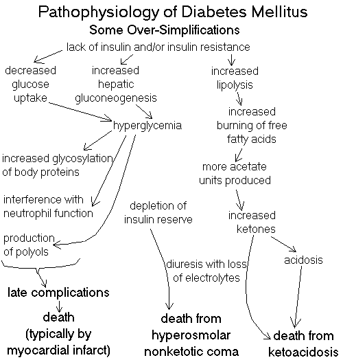
CLASSIFYING DIABETES MELLITUS AND RELATED CONDITIONS
![]() Diabetes Mellitus, pancreas
Diabetes Mellitus, pancreas
Text and photomicrographs. Nice.
Human Pathology Digital Image Gallery
| "Diabetes" literally means "siphon", because of the osmotic
diuresis produced by the glycosuria. This was known all-too-well to Hippocrates,
who may have named it.
Diabetes mellitus (MELL-uh-tuss, please) is "a chronic disorder of carbohydrate, fat, and protein metabolism characterized in its fully expressed clinical form by an absolute or relative insulin deficiency, fasting hyperglycemia, glycosuria, and a striking tendency toward the development of atherosclerosis, microangiopathy, nephropathy, and neuropathy" (old Big Robbins). |  |
Diabetes is our commonest serious metabolic disease, affecting maybe 5% of the US population. On the average, it takes 15 years off the patient's life (JAMA 285: 628, 2001) and accounts for a tremendous amount of health care expenses.
Worldwide, there were 171 million diabetics at the turn of the century. In 2008, the best estimate was 250 million (Lancet 371: 5, 2008). Expect at least 350 million by 2030 (Nature 444: 840, 2006; 380 million Lancet 371: 1723, 2008). You will need to know the terminology (which is often not used correctly):
Diabetes mellitus ("overt diabetes", "manifest diabetes", etc.): the patient has...
Two new terms were introduced early in the 2000's: "impaired fasting glucose" (IFG, i.e., 110-125 mg/dL) and "impaired glucose tolerance" (IGT, i.e., 121-179 mg/dL at the two-hour mark). See Arch. Int. Med. 161: 397, 2001. Clinicians are now using them, and finding that 10-15% of US adults have one of these (Am. Fam. Phys. 69: 1961, 2004.) The value of this information, if any, is unknown.
Type I diabetes and Type II diabetes (below) are sometimes called "primary diabetes", since they seem to be genetic diseases in their own right.
Secondary diabetes is said to exist when the metabolic disturbances are the result of some other identifiable illness, injury, molecular abnormality, etc., etc.
No one knows quite where to put "lipoatrophic diabetes", with severe lipodystrophy along with insulin resistance and type II diabetes. A few genes are known (J. Clin. Endo. Metab. 91: 2689, 2006; "perleptin" NEJM 364: 740, 2011).
Impaired glucose tolerance ("glucose intolerance", "subclinical diabetes", "asymptomatic diabetes", "chemical diabetes", "latent diabetes"): fasting blood sugar is normal, but a glucose tolerance test is abnormal, with a 2 hour peak between 140 to 199. These people are likely to go on to get type II diabetes. Looking for this was a fad in the early 1990's but probably did no one any real good: Am. J. Med. 105(1A): 15S, 1998.
Gestational diabetes mellitus: diabetes mellitus first appearing during pregnancy, and perhaps disappearing when the pregnancy ends. It's caused by the altered hormonal milieu.
By contrast "true gestational diabetes", which ends with the pregnancy, is "controversial." Mom's genetics and lifestyle both come into play. Babies are likely to be large; they may or may not really have more problems as newborns; supposedly they grow up to be fat kids and early type II diabetics but how much of this is due to genetics and family lifestyle is impossible to sort out. Further, the standard for diagnosis of "gestational diabetes" is the hated glucose tolerance test, and the research community doesn't accept fasting blood sugar levels as valid. The burning question is "Who do we screen to see if we need to put her on insulin?" It remains unanswered. This is awkward. See Lancet 373: 3789, 2009.
"Previous Abnormality of Glucose Tolerance" ("prediabetes", "latent diabetes"): the patient once had measurable glucose intolerance (as, when she was pregnant), but is chemically normal now (but may be at risk for future diabetes mellitus, depending on the circumstances).
"Potential Abnormality of Glucose Tolerance" ("prediabetes"): the monozygotic twin of a type II diabetic, or (less justifiably) someone else with a strong family history.
Not diabetes: Glucose intolerance only under some obvious physiologic stress (myocardial infarction, pneumonia, severe burns, terror of venipuncture, etc.) Mostly an epinephrine effect; probably cortisol contributes as well.
Type II: defects in insulin secretion, and/or a relative lack of insulin, and/or insulin resistance;
Neither: Diabetes due to damage to the whole pancreas (old pancreatitis, cystic fibrosis (tiny pancreases -- Br. J. Rad. 83: 921, 2010), hemochromatosis, cancer -- see below) and/or glucose intolerance from an endocrinopathy. Diabetes from a lipodystrophy (Am. J. Med. 108: 143, 2000), genetic or from HIV drugs, is now very familiar. I learned to call these "secondary diabetes". Neither: Gestational diabetes
A 1999 proposal by WHO and the American Diabetes Association (Br. Med. J. 317: 359, 1998) to use the term "type III diabetes" for disease of the whole pancreas or (for some reason) the simple-autosomal dominant forms of type II diabetes, and "type IV diabetes" for gestational diabetes, didn't catch on.
![]() Insulitis
Insulitis
Type I diabetes
WebPath photo
PRIMARY DIABETES TYPE I ("juvenile onset", "labile", "ketoacidosis-prone", "insulin-dependent"): 10% of diabetics (review Lancet 383: 69, 2014)
One person in 300 in the U.S. gets this kind of diabetes (rates vary considerably from nation to nation; * rates are higher at higher latitudes).
Typical case: A child (average age twelve years, but we now know you can get the disease at any age) presents with polyuria, polydipsia, and polyphagia of relatively sudden onset. The child is found to have very high blood glucose levels causing osmotic diuresis.
Before the era of injectable insulin, diabetic ketoacidosis (DKA) and death followed in short order.
You remember the pathophysiology of ketoacidosis from your physiology course. Future clinicians: Ketoacids impart the familiar "rotten apples" sweetness to these patients' breath.
Today, the child looks forward to a period of fairly good health while taking injectable insulin, checking blood glucose several times a day with chemical strips and a reflectance meter.
After 10-15 years, unless control is good, the diabetic starts to suffer with infections, eye problems, peripheral neuropathy, gangrene of the lower extremities, kidney disease, stroke, and coronary atherosclerosis.
Historically, death usually came about forty years after onset as the result of a myocardial infarction. By this time, 50% of patients had lost their kidneys, and nearly as many were blind, stroked out, legless, and/or in chronic pain from neuropathy. A well-treated, compliant diabetic typically does better today.
The essential lesion in type I diabetes is a severe absolute lack of insulin.
* Only half of patients have any evidence of insulin production (measure C-peptide in serum).
Insulin deficiency and hyperglycemia explain the presentation but do not explain the later complications of the disease.
"Type I diabetes is a genetically programmed, chronic autoimmune disease" (NEJM 314: 1360, 1986, an early review; update Nature 351: 519, 1991), with the acute-symptomatic phase sometimes triggered by an acute viral illness.
Genetic factors:
Siblings of those with Type I diabetes are at increased risk (25x).
Identical twins of those with Type I diabetes have maybe a 30% chance of eventually getting it also.
Type I diabetes is strongly associated with HLA-related antigens DR3 and DR4. (* If one has the misfortune to have both, it's even worse.... The former association with some HLA-B antigens was due to their linkage to DR3 and DR4; and currently, it appears that the also-linked DQ is the closest important site.)
* As is so common when the immune system attacks gland parenchyma, the beta cells of these patients express HLA class II histocompatibility antigens. No one knows whether this is cause or effect.
* The molecular defect that permits type I diabetes to occur seems to be homozygous absence of aspartic acid in position 57 of the HLA class II DQ chain (Nature 329: 599, 1987; Nature 333: 710, 1988), at least in the U.S. Update on HLA links: J. Clin. Endo. Metab. 89: 4037, 2004; they vary tremendously from nation to nation: J. Clin. Endo. Metab. 90: 5104, 2005.
The famous locus IDDM1, where certain polymorphisms give a risk for type I diabetes, is a component of the HLA system (Diabetes 50: 1200, 2001).
The gene IDDM2 ("implicated in diabetes mellitus") is a complicated, highly variable tandem repeat adjacent to the real insulin gene. Two variants are strongly linked to type I diabetes (update Diabetes 53: 1884, 2004).
The classic animal model of autoimmune diabetes is the non-obese diabetic (NOD) mouse, which gets that way because of genes at three (or more) loci (Nature 353: 260, 1991; J. Imm. 152: 204, 1994). Updates Nat. Med. 5: 601, 1999; J. Immuno. 169: 6617, 2002; to date; the exact reasons for the famous mouse's diabetes remain elusive, though they involve altered T-cell function.
* The BB (formerly BB/W) rat is a strain discovered in 1977. These rats have autoimmune insulitis, and the majority develop acute-onset type I diabetes. They helped us find the IDDM1 and IDDM2 loci (Acta. Diabet. 35: 109, 1998).
Autoimmune factors:
Several types of IgG anti-beta-cell antibodies occur. One or more is present in the vast majority of type I diabetics the acute phase (contrast 0.5% in healthy people). It is now quite clear that they are etiologic, and that they are usually present before age 2 in children destined to get type I diabetes (Ann. Int. Med. 140: 882, 2004); most (but not all) youngsters with these autoantibodies will eventually develop diabetes (JAMA 309: 2473, 2013).
It's now clear that there is an early (first year of life) antibody response directed against insulin itself -- an attempt to tolerize these children failed to decrease the risk of ensuing diabetes, but stay tuned (Lancet 372: 1746, 2008).
Other autoantibody specificities include anti-glutamic acid decarboxylase (Nature 347: 151, 1990; NEJM 322: 1555, 1990; Lancet 341: 1378 & 1383, 1993; diabetogenic epitope Lancet 343: 1607, 1994; true both of NOD mice and people: Nature 366: 69 & 72, 1993).
There is also cell-mediated immunity directed against beta cells in most patients who have been studied. Again, one autoantigen is glutamic acid decarboxylase. Update Nature 391: 177, 1998.
There was generally a dense lymphocytic infiltrate in the islets of patients dying in the acute phase (such deaths are thankfully very rare nowadays).
* Maybe 1 in 5 of these people ends up with another autoimmune glandular disease (autoimmune Addison's disease, Hasmimoto's autoimmune thyroiditis, Grave's disease of the thyroid). Likewise, plenty of people, with or without other autoantibodies, have anti-islet cell antibodies but never go on to develop autoimmune diabetes. There are now three known syndromes, each with a genetic link to immune-regulating genes (J. Clin. Endo. Metab. 91: 1210, 2006).
Type I diabetes has been increasing in frequency for the past several decades. No one knows why, and there is a massive, amazingly inconsistent literature looking for possible causes (Lancet 371: 1730 & 1777, 2008).
* The only one of dozens of possible explanations for the increase that I'll trouble you with is the one about cow's milk.
All the recent stuff is from obvious "independent thinkers" (ignoring what we know of immunology: Food & Chem. Tox. 42: 707, 2004) and studies that invited recall bias (Ann. Nutr. Metab. 47: 267, 2003).
The non-obese diabetic mouse does get some protection from drinking mother's milk instead of cow's milk. The experimentalists speculate at length about how perhaps this is because mother's milk contains insulin and/or other peptides to which the gut lymphocytes need to become tolerant (Diabetes 48: 1501, 1999). But think -- the experiment requires taking the experimental mice away from their mothers. This must have many far-reaching effects beyond just the exposure to cow's milk.
The TRIGR of hydrolyzed infant formula (i.e., no antigens) as a way of preventing type I diabetes was a complete and utter failure. This tells me that diet isn't the cause of type I diabetes. JAMA 311: 2279, 2014.
Viral factors: Clinically, Type I diabetes often follows a viral illness.
Worth knowing: Kilham rat parvovirus infection produces type I autoimmune diabetes in diabetes-resistant rats (Diabetes 45: 557, 1996; J. Immuno. 165: 2866, 2000). This is now a robust finding (J. Imm. 173: 137, 2004; J. Imm. 178: 693, 2007).
* Interest in another virus (retrovirus IDDMK(1,2)22), which was said to act as a superantigen, seems to have faded.
A Coxsackie B4![]() virus from the
pancreas of a patient dying shortly after the onset of the illness
destroys the beta cells of NOD (non-obese diabetic) mice; it's now clear that the virus causes a chronic infection of these
islands
(J. Inf. Dis. 171: 1131, 1995). Since this article, Coxsackie CB4
has been found commonly as a recent infection in kids coming down with
diabetes. The most impressive work I've seen is from Italy, where three of six
type I diabetes and none of 26 controls had coxsackie B4 in their islets
(Proc. Nat. Acad. Sci. 104: 5115, 2007).
virus from the
pancreas of a patient dying shortly after the onset of the illness
destroys the beta cells of NOD (non-obese diabetic) mice; it's now clear that the virus causes a chronic infection of these
islands
(J. Inf. Dis. 171: 1131, 1995). Since this article, Coxsackie CB4
has been found commonly as a recent infection in kids coming down with
diabetes. The most impressive work I've seen is from Italy, where three of six
type I diabetes and none of 26 controls had coxsackie B4 in their islets
(Proc. Nat. Acad. Sci. 104: 5115, 2007).
The mechanism of Coxsackie B4 induction of diabetes now seems clear -- the NOD mouse has lots of autoreactive-but-unactivated T-cells, Coxsackie B4 produces a mild infection of the beta cells, and bystander T-cells are activated ("bystander activation"). Happens in mice and maybe in kids.
* The whole business has become enormously complicated, with the randomness of the T-cell system interacting with a host of genes (J. Immuno. 188: 294, 2012; J. Immuno. 189: 1406, 2012).
Overwhelming infections with mumps![]() or cytomegalovirus
or cytomegalovirus![]() also
have been implicated in rare cases of "type I diabetes". The pancreas
can be destroyed by congenital rubella
also
have been implicated in rare cases of "type I diabetes". The pancreas
can be destroyed by congenital rubella![]() .
.
A huge search for the "insulitis virus" in humans using molecular probes found nothing: JAMA 257: 1145, 1987. Probably this is still good. Enterovirus is "usual suspect" as trigger for autoimmunity, and perhaps there's something to it (BMJ 342: d35, 2011).
What does all this mean? In most cases of type I diabetes, it is hypothesized that a viral infection triggers autoimmune destruction of the beta cells in genetically-predisposed individuals.
However, in most of these children, there have been progressive abnormalities of glucose metabolism in these patients long before the onset of illness (Br. Med. J. 294: 5, 1987).
Some stress ("maybe the virus") apparently causes decompensation at the "time of onset". Following recovery from the first episode of ketoacidosis, the "honeymoon period" begins, when control is easy for several years. (* Patients continue to produce some of their own insulin -- i.e., there is C-peptide in their blood -- during the "honeymoon".)
* No one knows why type I diabetes is becoming more common, but there's no question that this is happening (Lancet 373: 1999, 2009). Studies that suggest a major, unknown environmental factor (Lancet 364: 1699, 2004) make me think we are dealing with a viral trigger.
PRIMARY DIABETES TYPE II ("adult onset", "stable", "ketosis-resistant", "non-insulin-dependent"): 90% of diabetics. Update Lancet 383: 1068, 2014.
Typical case:
An overweight adult (most over age forty) is discovered on routine screening to have elevated fasting glucose or glycosuria.
In other cases, the diabetes is discovered during evaluation of impotence, pain, eye trouble, stroke, foot trouble, bad infection, or coronary disease.
Some patients have their diabetic predisposition unmasked by pregnancy. Such women get better after the pregnancy, but are at greater risk for eventually developing type II diabetes.
Before the era of injectable insulin, nothing much was done for type II diabetics, even if the disease was detected. The patients got complications and had shorter life spans.
Today, the adult looks forward to dieting, doing aerobic exercise, and possibly getting treated with insulin or "diabetes pills", probably getting an ACE inhibitor, and maybe a statin for lipid control. Complications will occur as in Type I diabetes, depending on how well the patient is able to manage the hyperglycemia. Death will probably be due to a myocardial infarct.
Type II diabetes is a polygenic disorder, with its expression modified by a person's exercise habits and amount of bodyfat.
Identical twins have nearly 100% concordance for Type II diabetes. There are no good HLA associations or phenomena pointing to autoimmunity.
It is now absolutely clear that the onset of type II diabetes is fairly easily delayed by lifestyle interventions, notably diet and exercise (Lancet 368: 1673, 2006). Current thinking about the common disease is "lipocentric", i.e., excess bodyfat generates insulin resistance, leading to high blood glucose and non-enzymatic glycosylation of the glucose regulators themselves, a vicious cycle (JAMA 299: 1185, 2008).
A subtype of diabetes ("not really type I or type II) that can present in young people (maturity-onset diabetes of the young, MODY) is an autosomal dominant with 90% penetrance, and several loci. Someday we may change the name to "single-gene diabetes." See below.
There is decreased but not absent insulin, and little or no insulin resistance. The name suggests that this is adult-onset / type II diabetes in a young person; this is not the case.
MODY accounts for about 10% of diabetics in some communities, and less-severe alleles of the genes are of course implicated in common type II diabetes.
The first defect to be discovered was in the glucokinase gene (Nature 356: 721, 1992; mechanisms Lancet 340: 444, 1990; diagnosis Lancet 345: 1313, 1995; pathophysiology Diabetes 46: 204, 1997; this enzyme, as you remember, is the key link in the signalling system by which beta cells monitor blood glucose).
There are more than eight MODY genes known today, all autosomal dominant, all in the insulin-release system (Proc. Nat. Acad. Sci. 94: 13209, 1997; Diabetes 47: 1459, 1998; Diabetes 52: 872, 2003). The most common may be "hepatocyte nuclear factor 1-alpha". Update on genes Diabetes 53: 1894, 2004.
A single major genetic defect at any of several type II diabetes locus, and/or several minor defects at several of the loci, seems to be the underlying cause of type II diabetes.
To date, we know almost nothing about how beta cells replicate and are replenished, especially in adults; since some of the genes have to do with the cell cycle, perhaps we'll find answers here -- or frustration when we "try to grow new beta cells in adult-onset diabetics" (Nat. CP Endo. Metab. 3: 758, 2007).
The Type II diabetes genes:
1%... mitochondrial DNA syndromes (often goes with deafness; Ann. Int. Med. 134: 721, 2001; many others)
?%... the mitochondrial uncoupling proteins (Diabetes 47: 1528, 1998; Diabetes 53: 1905, 2004).
1%... glucokinase
1%... insulin itself
1%... insulin receptor (* the severe form is "leprechaunism", a "progeria": Biochim. Biophys. Acta.1402: 86, 1998)
15%... insulin receptor substrate (IRS-1, it's very complicated and results are mixed: Diabetes 52: 1544, 2003; J. Clin.Inv. 114: 908, 2004)
1%... GLUT4, the glucose-through-the-membrane transporter
?%... adiponectin, released from adipocytes, causes liver and muscle to burn triglyceride and be more insulin-sensitive (Nat. Med. 7 887, 2001; Nat. Med. 10: 452 & 524, 2004; Diabetes 53: 1150, 2004).
?... adiponectin receptors 1 and 2 (Diabetes 53: 2132, 2004; Diabetes 54: 2245, 2005)
?%... hepatocyte nuclear factor alpha (HNF-1alpha/TCF1, causes MODY3; risk for classic type II; Diabetes 53: 2122, 2004; Diabetes 54: 2336, 2005; Diabetes 53: 1141 & 3002, 2004); also TCF7L2 (NEJM 355: 241, 2006; Nat. Genet. 39: 218, 2007; Nat. Genet. 39: 218, 2007)
?%... ICAM-1 (Lancet 362: 1723, 2003)
?%... calpain 10 (J. Clin. Endo. Metab. 87: 2606, 2002)
?%... beta adrenergic receptors (gives the munchies / obesity and diabetes: NEJM 333: 382, 1995; Clin. Endo. 59: 476, 2003).
?%... leptin (must be rare in humans, though of course in mice it's famous as the ob/ob model for type II diabetes)
?%... leptin receptor (again, probably rare in humans, though of course in mice it's famous as the db/db/ model for type II diabetes) ?%... phosphoenolpyruvate carboxykinase (J. Clin. Endo. Metab. 89: 898, 2004.
?%... Sulfonylurea receptor (Lancet 361: 22, 2003).
?%... HIF-1alpha (induces VEGF, J. Clin. Endo. Metab. 90: 5841, 2005).
?%... alpha2-Heremans-Schmid glycoprotein (Diabetes 54: 2477, 2005)
?%... ABCC8, the ATP-sensitive potassium channel at the beta-cell sulfonylurea receptor; mutations here cause neonatal diabetes (NEJM 355: 456, 2006)
?%... mitochondrial fat-burning systems (no gene yet; NEJM 350: 664, 2004);
?%... mitochondrial leucyl tRNA synthetase / LARS2 (Diabetes 54: 1892, 2005)
?%... sterol regulatory element binding protein (SREBP)-1 (Diabetes 53: 842 & 2153, 2004
?%... K(ATP) channels in muscle (?!; Diabetes 54: 1592, 2005)
?%... Wolfram syndrome locus (WFS1; Nat. Genet. 39: 951, 2007)
?%... CDKAL1 (Nat. Genet. 39: 770, 2007
?%... Alpha2A adrenergic receptors, overexpressed (treat with yohimbine?!): Science 327: 217, 2010)
?%... Vav3 -- a new gene for non-obese mice who get autoimmune diabetes (J. Immuno. 184: 5075, 2010.)
lots of variants especially in white folks -- HMGA1 (JAMA 305: 903, 2011)
* The lipoatrophic diabetes mouse (knockout at two loci) has zero bodyfat and extreme insulin resistance with diabetes. This is a model for both a few human genetic syndromes and the lipoatrophy of HIV patients on protease inhibitors (Ann. Int. Med. 133: 304, 2000).
Type II diabetes is now rampant in the developing world and many of our own First American peoples.
Until recently, the tendency was to blame the western diet ("the poor nations have been coca-colonized": Nature 357: 362, 1992). I have always taught that the real reason is that the world's poor are much better-fed than in the past, and most no longer lead lives of constant hard physical labor. Stay tuned.
Of course, there has been stronger natural selection against diabetes in countries like the U.S. and Western Europe that have been well-fed for centuries. And in societies with episodes of famine, there is a strong selection bias for type II diabetic body chemistry (i.e., a tendency to hang onto carbohydrate calories), and little chance to express the phenotype.
Whether or not it's related, unborn children exposed to famine have a much stronger tendency to develop type II diabetes when they grow up: Lancet 351: 173, 1998; this is now a robust finding Diabetes 61: 2255, 2012. The same is probably true of children exposed to famine after birth (Diabetes 61: 2255, 2012).
* The Pima Indians present a special problem; their rate of diabetes is extremely high with a host of different genetic mutations for insulin resistance (update Diabetes 53: 1181, 2004).
By age 65, the following percentages of U.S. ethnic groups have diabetes:
Hispanics 33%
Blacks 25%
Whites 17%
The pathophysiology of type II diabetes is fairly well understood.
In type II diabetes, basal insulin secretion is generally normal. In response to glucose administration, insulin secretion may be abnormally low, normal (rare), abnormally high, or delayed ("too much, too late").
Most Type II diabetics have insulin resistance in both liver and skeletal muscle, and this appears to be the key lesion. In addition, however, there is almost always some evidence of beta cell dysfunction.
The liver continues to make and put out glucose (gluconeogenesis) when blood sugar is high, and fails to take up orally-administered glucose. The skeletal muscles fail to take up glucose in response to insulin.
The amount of insulin resistance is modified by obesity and physical conditioning. There's also the baffling combination of gut polypeptides, prostaglandins, beta-endorphins, etc., etc....
Exactly how obesity itself increases insulin resistance is enormously complex. Update Nature 444: 840, 2006.
You already know the metabolic syndrome / metabolic syndrome X (truncal obesity, insulin resistance, dyslipidemia). The cause remains obscure.
One suspect is resistin, produced by adipocytes, and able to render muscle and liver resistant to the effects of insulin (NEJM 345: 1345, 2001). It's produced especially well by the abdominal and omental adipocytes; this may explain the special risk of "central obesity".
* A locus for metabolic syndrome DYRK1B: NEJM 370: 1909, 2014.
* Hormone sensitive lipase (LIPE), even with one null allele, confers the metabolic syndrome and type II diabetes NEJM 370: 2307, 2014.
It's taken time, but pathologists are finally backing up what clinicians have been saying about the "inflammatory phenotype". Whether or not this is the best choice of words, we now know that the bellyfat of people with diabetes and/or the other features of the metabolic syndrome (though NOT the fat of equally-fat people with normal glucose tolerance) is
Remember that there are plenty of fat kids and teens with the metabolic syndrome (Lancet 369: 2059, 2007).
* Liposuction completely fails to alter the metabolic abnormalities caused by obesity (NEJM 350: 2549, 2004).
Two other players in the complex business of insulin resistance is a pair of little-known hormones, amylin ("islet-amyloid polypeptide", "IAPP", pumped out of beta cells along with insulin) and * calcitonin-gene related polypeptide (CGRP, h-CGRP, from nerve and gut), both acting on skeletal muscle to increase its resistance to insulin (PNAS 88: 7713, 1988). They act on the same receptor, which is not present in fat or parenchymal cells (Diabetes 40: 395, 1991; Diabetes 40(S1): #267, #255, several others, 1991).
Of course, amylin is absent in type I diabetics. Amylin has been reported to be greatly increased in the serum of some type II diabetics. Excreted through the kidneys, it also might account for some of the insulin resistance in renal failure. See Diabetes Care 39(S1): A111-A113, 1990.
Disregarded for many years after its discovery, new work re-emphasizes amylin as an important player in type II diabetes (J. Clin. Endo. Metab. 89: 3629, 2004. Pramlintide is a synthetic amylin analogue (Diabetes 53-S3: S-233, 2004; Denmark) that's now approved by the FDA as an adjunct. Injected at mealtime, it seems helpful in both type I diabetes ("the first medicine to lower glucose in type I diabetes since insulin) and type II diabetes.
* IAPP pieces ("cylindrins") inside beta cells as possible mediators of beta-cell loss: Diabetes 62: 327, 2013.
As a matter of fact, the whole business of fat in diabetes has just gotten complicated by the observation, using the latest MRI technology of course, that bodyfat distribution in type II diabetics differs considerably from fat non-diabetics. Diabetics have less subcutaneous fat, and more visceral and (surprise!) intramuscular fat (Am. J. Clin. Nutr. 89: 807, 2009.) Prediabetic (fasting glucose 100-125 mg/dL or 2-hour glucose 140-199 mg/dL) teens have more visceral, hepatic and pancreatic fat (J. Clin. Endo. Metab. 98: 1115, 2013).
I predict that when the underlying cause of type II diabetes (i.e., simultaneous insulin resistance and aberrant insulin production) is worked out, it will prove to be primarily a mitochondriopathy. Stay tuned.
The "Somogyi phenomenon" is a rebound hyperglycemia from all the stress hormones that pour out when the blood glucose drops too low from too much insulin. If a diabetic is hungry, gaining weight, and feeling crummy, consider reducing the insulin levels.
The "dawn phenomenon, i.e., hyperglycemia and insulin resistance in the morning without previous "Somogyi" hypoglycemia, is due to the high output of hGH during while you're finishing up your sleep in the morning.
* WARNING: Many clinicians use the term "insulin resistance" to refer instead to hard-to-manage diabetics of any type who require more than 200 units of insulin daily (a whopping dose). Many of these patients have antibodies against insulin, while others have severe type II diabetes or any of several other problems. See NEJM 315: 212, 1987.
Hyperosmolar nonketotic diabetic coma (HNKK, HONK) is the usual cause of "diabetic coma" in Type II diabetics (see Arch. Int. Med. 147: 499, 1987), though most of them never get it.
Classically, some acute stress (often the 'flu) increases the demand on the Type II diabetic's struggling beta cells, and the supply of insulin is exhausted. Plasma glucose levels suddenly go extremely high, causing osmotic diuresis, electrolyte disturbances, and death.
Or the illness may simply cause dehydration, producing a vicious cycle with insulin resistance, stress hormones, soaring glucose levels, and ongoing dehydration.
Ketoacidosis is uncommon in type II diabetes, but can occur.
SECONDARY DIABETES has many etiologies
Pancreatic diabetes: destruction of the islets by disease of the exocrine pancreas.
Causes: pancreatitis, carcinoma, hemochromatosis ("bronze diabetes" -- don't overlook this one!), trauma, surgery, etc. etc. Diabetes from cystic fibrosis is uncommon but happens J. Ped. 142: 97, 2003)
Endocrine diabetes: glucose intolerance due to other endocrine disturbances
Causes: Cushing's syndrome (from any cause), acromegaly, amylin from pancreatic cancer, obesity (??), stress, amylin production by cancer of the pancreas (see above), etc. etc. Glucose intolerance from the lipodystrophies / anti-retroviral therapy will end up here, especially after all the hormones involves get sorted out. It would be logical to place pregnancy here too, though it is officially classed elsewhere.
Some people put the one-gene insulin resistance syndromes here, which makes less and less sense every year as these genes turn out to be alleles for standard Type II diabetes.
Rarely, people make autoantibodies that block insulin receptors (South. Med. J. 92: 717, 1999). Update J. Clin. Endo. Metab. 89: 2222, 2004; contrary to popular belief, acanthosis nigricans in a young diabetic (while commonly seen) does not imply antibodies to insulin receptors.
REMEMBER: Regardless of the cause of the prolonged hyperglycemia, we now know that the complications in remote organs (arteries, eyes, kidneys, nerves) will be the same.
ANATOMIC PATHOLOGY OF DIABETES MELLITUS: These are usually the effects, rather than the causes, of hyperglycemia.
DIABETIC BLOOD VESSEL DISEASE
LARGE VESSEL DISEASE ("macroangiopathy"): accelerated atherosclerosis
Diabetics have a variety of poorly-understood disturbances of lipid metabolism. Nonenzymatic glycosylation of lipoproteins seems to be a problem, LDL's stick best to glycosylated collagen, etc., and glycation products (when they bind to their special receptors in the intima) cause the production of fibrous tissue.
The result is the rapid development of severe atherosclerosis, with strokes, gangrene of the lower extremities, and myocardial infarcts taking their toll, often early in life. Of course, this is all much worse if the diabetic also smokes cigarets.
{09378} diabetic gangrene
{48076} diabetic gangrene
{48022} diabetic ulcer
{48023} diabetic ulcer
{48150} diabetic ulcer
SMALL VESSEL DISEASE ("microangiopathy"): hyaline arteriolar sclerosis
This is a complex problem that is attributed primarily to advanced glycation end products.
The basement membrane of the capillaries and the arterioles becomes much thicker ("hyaline arteriolar sclerosis"). Its expansion eventually compromises the lumen of the vessels.
Not surprisingly, these vessels are relatively inelastic, and this is an early, important problem: Br. Med. J. 312: 744, 1996.
Even if the lumen is not badly compromised and the wall isn't excessively stiff, the small vessels of diabetics open and close chaotically, and proper tissue perfusion cannot be assured.
Additionally, the pericytes can proliferate (especially in the glomeruli, where pericytes are called "mesangial cells") or die off (especially in the retina, where pericytes are called "mural cells"). This causes trouble at both sites.
* Endothelial cells can also proliferate, narrowing the lumen further.
* Other factors that are cited are the over-sticky platelets of diabetics, increased blood viscosity, increased RBC rigidity, and increased numbers of free radicals.
Diabetic microangiopathy in the hepatic sinusoids (the newly-recognized "diabetic hepatosclerosis"): Arch. Path. Lab. Med. 130: 27, 2006; Am. J. Clin. Path. 132: 494, 2009.) Basement membrane is thickly deposited along the sinusoids (* future pathologists: stain with laminin).
Microangiopathy hugely augments the ischemia caused by atherosclerosis, which is why so many diabetics lose legs. It may account for other problems also.
Yes! Tight diabetic control reduces and even reverses microangiopathy. See NEJM 309: 1546 & 1551, 1983, and many others since.
Most diabetics eventually become hypertensive as a result of inability to handle a sodium load (Am. J. Med. Sci. 307(S1): S-53, 1994.) No one knows for sure why, but it seems reasonable that the narrowing of the small arteries in the kidney is at least part of the problem.
Many diabetics are greatly troubled by congestive heart failure as the disease progresses, and perhaps nonenzymatic glycosylation of the heart muscle proteins itself is part of the problem, since even if you control for other factors, poor glycemic control correlates strongly with the development of CHF (Circulation 103 2668, 2001).
A massive study of brains of older folks with dementia revealed how commonly older diabetics (who are at greater risk for dementia) have huge numbers of micro-infarcts without very much Alzheimer-type pathology. Not surprising, but beautifully documented (Arch. Neuro. 66: 315, 2009; correlates with cognitive decline of course Neurology 75: 997, 2010. Hyperglycemia and insulin resistance as factors for the development of Alzheimer neuritic plaques: Neurology 75: 74, 2010. Problems with the white matter independent of vascular damage load: Diabetes 62: 2112, 2013.
DIABETIC KIDNEY DISEASE ("diabetic nephropathy"; Disease-A-Month 44: 214, 1998; NEJM 341: 1127, 1999):
Renal failure causes much disability and death among type I diabetics; this is now the #1 single cause of end-stage renal disease in the U.S. Type II diabetics generally die of something else before their kidneys fail.
Renal vascular lesions
Arteriolar sclerosis of both afferent and efferent arterioles at the glomerular pole is highly characteristic of diabetes. (The other diseases of renal arterioles, notably common-type high blood pressure, only cause sclerosis of the afferent arteriole.)
* Atherosclerosis of intrarenal arteries is common in diabetics and rare in non-diabetics; it is not the major problem.
Glomerular lesions
Always present:
1. Thickening of the glomerular basement membrane because of increased production of GBM (sometimes called "diffuse glomerulosclerosis").
2. Increased amounts of mesangial matrix (also sometimes called "diffuse glomerulosclerosis"). Increased number of mesangial cells in the early lesion, later decreased as the entire glomerulus is replaced by matrix ("hyalinization" of the glomerulus.)
* 3. The GBM, mesangial matrix, and tubular basement membranes (also thick) bind albumin and other proteins non-specifically ("all that sticky sugar....")
* These three features, together, are pathognomonic of diabetes mellitus (but you probably knew already....) They occur separately in other diseases.
Often present:
Nodular glomerulosclerosis or (nodular) Kimmelstiel-Wilson disease. Big balls of GBM-mesangial matrix material in the glomerular tufts. Highly characteristic of diabetes or FSGS.
{08892} KW disease; note balls of hyaline, and thick GBM
(i.e., you can actually tell where it is)
{17159} diabetes with hyalinized arteriole
{16789} diabetic glomerulosclerosis, electron micrograph (thick GBM)
{16790} diabetic glomerulosclerosis, electron micrograph (thick GBM)
{16791} diabetic glomerulosclerosis, H&E
{16792} diabetic glomerulosclerosis, PAS; nice
capsular drop too
{16793} diabetic glomerulosclerosis, H&E
{08893} Kimmelstiel-Wilson diabetic nodular glomerulosclerosis;
H&E
{08895} Kimmelstiel-Wilson diabetic nodular glomerulosclerosis, PAS
{09877} Kimmelstiel-Wilson diabetic nodular glomerulosclerosis
{17158} Kimmelstiel-Wilson diabetic nodular glomerulosclerosis
{17171} end-stage diabetic glomerulosclerosis
|
|
|
Sometimes present:
* "Fibrin caps" ("exudative lesion", "hyperfiltration lesion") -- hyaline frosting on a glomerular tuft
* "Capsular drops" -- hyaline material on the inside surface of Bowman's capsule (highly characteristic of diabetes.)
Clinically, patients have albuminuria (rarely heavy proteinuria), then renal failure (probably due to the mesangium crunching the glomerular capillaries).
The etiology of diabetic glomerulopathy is complex and poorly-understood. Intrarenal fluid dynamics are involved. We don't even know why the kidneys enlarge in diabetics (NEJM 324: 1662, 1991, still good).
Tight control of blood glucose does seem to benefit these patients, and reduces the hyperfiltration response to amino acids (NEJM 324: 1629, 1991). Patients are now put on ACE-inhibitors and protein-restricted to prevent progression of the renal disease. (Yes, it can regress due to therapy: NEJM 348: 2285, 2003).
* Ace-inhibitor plus a calcium channel blocker works marvellously to prevent diabetic kidney disease: NEJM 351: 1941, 2004.
Other renal lesions in diabetes:
Thick tubular basement membranes (not a health problem).
* Fatty change of tubular cells (systemic lipid disturbance, not a health problem).
Glycogen in proximal tubular cells (Armanni-Ebstein lesion, a sign of heavy glycosuria, not itself a health problem).
{46306} Armanni-Ebstein; lots of glycogen in the tubular cells
Kidney infections (gram-negative bacilli causing infection of renal pelvis in pyelonephritis,
staphylococci![]() causing cortical infections,
candida
causing cortical infections,
candida![]() infections, etc.)
infections, etc.)
Renal papillary necrosis -- just like it sounds. The lesion is seen in diabetes, obstruction, sicklers, Wegener's, or abuse of the analgesic phenacetin.)
{49306} pyelonephritis and papillary necrosis in a diabetic
EYES: Diabetes is the commonest cause of blindness before old age in the US. Classic review: Lancet 350: 197, 1998.
Cataracts: a variety of types, including some clearly caused by sorbitol deposition (proof Proc. Nat. Acad. Sci. 9: 2780, 1995; remains a robust finding).
Glaucoma: reason for its being more common with diabetes is uncertain.
Diabetic retinopathy: the most serious diabetic eye problem (Lancet 376: 124, 2010)
Nonproliferative phase (NEJM 322: 978, 1990)
Edema (especially of the macula NEJM 372: 1193, 2015), protein exudates, lipid deposits, hemorrhages, microinfarcts ("cotton-wool patches") all indicate vascular problems
Microaneurysms (the first change, and highly characteristic of diabetes): ballooning of capillaries where perhaps a pericyte has come off.
{09365} diabetic retinopathy; hemorrhages and exudates
{22036} diabetic retinopathy; microaneurysms
{22039} diabetic retinopathy
{22042} diabetic retinopathy, notice the hemorrhages
{22045} diabetic retinopathy -- bleed
{22904} microaneurysm
{23156} cotton wool patches
{23180} diabetic retinopathy
{23183} diabetic retinopathy
Proliferative phase: new vessels grow, eventually invading vitreous humor, with hemorrhage, granulation tissue, fibrosis, retinal detachment. These patients get photocoagulation.
{09366} proliferative retinopathy
{22895} proliferative retinopathy
{22901} proliferative retinopathy -- "scar contracts"
and tears off the retina
The molecular biology is starting to be sorted out, with VEGF inhibitors now standard and producing obvious improvement in severe disease (NEJM 366: 1227, 2012). Sudden normalization of a poorly-controlled diabetic's glucose can accelerate proliferative retinopathy (Arch. Ophth. 116: 874, 1998).
PERIPHERAL NERVES (morphology: Diabetes 46 S 2: S 50, 1997)
Manifests as symmetrical sensory loss, sometimes with uncomfortable paresthesias, and as autonomic disturbances such as diarrhea (Am. J. Gastro. 94: 2165, 1999), bladder problems, orthostatic hypotension, impotence. Less often a mononeuropathy, perhaps due to infarction of a nerve.
Axons are lost, and Schwann cells also take a beating.
Some pathologists are now biopsying the gastric mucosa and counting the nerve twigs, which are much decreased when diabetic autonomic neuropathy is present: Neurology 75: 973, 2010.
{48174} Charcot's neuropathic joint changes
Probable chemistry: increased availability of glucose for polyol pathway results in more sorbitol.
* Aldose reductase produces polyols that are linked to the late complications in nerve and kidney. Inhibitors were not a great success for the neuropathy, but different aldose reductase alleles confer susceptibility to or protection from the glomerulopathy. (Diabetes 46: 287, 1997.)
The most interesting recent work in diabetic neuropathy focuses on the ability of ACE inhibitors to stop the progression independent of effects on blood pressure (NEJM 345: 851, 2001; works in rats too Diabetes 55: 341, 2006).
PANCREATIC ISLETS -- except when the islands are actually destroyed, the relationship of morphologic lesions to the abnormal metabolic state is usually obscure.
In Type I diabetes mellitus, early cases show lymphocytic infiltration ("insulitis"). Later cases show destruction of beta cells and even whole islets.
Degranulated and/or glycogen-filled beta cells have also been described.
In Type II, the islets most often look normal.
In either Type I or Type II, there may be hyalinization of the islets. In type I, it's collagen of course; in type II, it's collagen and/or amyloid. The latter is beta-pleated amylin, that little-known islet hormone. It is common in Type II diabetes, it may precede the overt disease, and it now appears that its massive accumulation in islets of type II diabetes does impair insulin production, and possibly is toxic to the beta-cells (Nature 368: 756, 1994). Confirmation: J. Clin. Endo. Metab. 89: 3629, 2004; Am. J. Path. 176: 861, 2010.
![]() Amyloid islet
Amyloid islet
Type II diabetes
WebPath photo
{08084} hyalinized pancreatic islets, type II diabetes
Babies of diabetic mothers have hyperplastic islets (because of all that glucose), and they are infiltrated with lymphocytes and eosinophils (mysterious.)
OTHER PROBLEMS FOR DIABETICS
Infections (bacterial and fungal)
Why diabetics get more infections is still poorly-understood.
Candida![]() may thrive on the glucose,
hyperglycemia slows down polys, poor circulation keeps the body from fighting infection, etc., etc.)
may thrive on the glucose,
hyperglycemia slows down polys, poor circulation keeps the body from fighting infection, etc., etc.)
*
{48090} diabetic abscess
{48091} diabetic abscess
Gallstones (made of cholesterol; nobody knows why these are more common in diabetics, but the average gallbladder volume is much higher in non-insulin-dependent diabetics, perhaps promoting stasis and nidation: Dig. Dis. Sci. 43: 344, 1998.)
Altered platelet function (significance?)
Complications of pregnancy -- all the common problems are commoner in diabetic mothers, and babies are bigger (partly the hyperglycemia, probably partly some growth factor or other: Br. J. Ob. Gyn. 103: 427, 1996) and at extra risk for a variety of birth defects (all of which seem to be preventable by euglycemia through pregnancy).
Diabetic xanthomas (yellow skin bumps -- pseudotumors made of lipid-laden macrophages), necrobiosis (focal necrosis of the dermis), and many other skin abnormalities
![]() Diabetic skin
Diabetic skin
Lecture notes and
some great photos
{12214} necrobiosis lipoidica diabeticorum
Hepatic fatty change, even in sober diabetics ("non-alcoholic steatohepatitis", which runs the gamut through neutrophils and Mallory's hyaline to micronodular cirrhosis). Of course this is all tied in with metabolic Syndrome X. Stay tuned.
* Scleredema -- pseudosclerodermatous changes over the back and shoulders caused by accumulation of glycosaminoglycans. This may be a marker for longstanding poor control.
* "Diabetic dermopathy" is purple-brown patches on the shins (less often, the upper legs and/or forearms) which may grow to coalesce. This supposedly has something to do with the microangiopathy and may be seen in other situations with vascular insufficiency.
Chronic hyperglycemia results in non-enzymatic glycosylation of many body proteins.
Hemoglobin A1c is glycosylated hemoglobin that can be measured in the blood to assess the quality of diabetic control (though, of course, home blood glucose testing several times a day by a highly motivated patient is even better....)
Estimaged average glucose (mg/dL) is 28.7 x A1c - 46.7.
There are numerous pitfalls to the HgbA1c test -- recently-transfused red cells are loaded from the glucose in the preservative, anemic / hemolyzing patients need the value adjusted, and people with variant hemoglobins are hard to assay.
Probably non-enzymatic glycosylation is part of the process that gets type II diabetes going and keeps it going, as a vicious cycle. Non-enzymatic glycosylation of the liver protein CRTC2, which stops glucose production by the liver in response to insulin, renders it unresponsive (Science 319: 1402, 2008). It probably also alters the function of beta cells (Transplantation 76: 657, 2003). There will be much more.
In the mid-1980's, your lecturer did a little work in residency on advanced glycation products in diabetic nephropathy, though without a paper. Since then, the field's exploded. "Advanced glycosylation (glycation) products" are proteins that have undergone a series of reactions with glucose.
Today, we can block their formation and break them up using alagebrium (formerly ALT-711). As a way of preventing diabetic complications, this seems promising (Endocrinology 148: 886, 2007). If this class of drugs comes into use, it'll be as "AGE inhibitors".
Hemoglobin A1c is an obvious choice for a diabetes screening device, and has been studied as such (JAMA 276: 1264, 1996); it's still not in common use.
* ADDITIONAL INFORMATION ABOUT DIABETES
|
One of the great stories from medical history is Banting and Best's visit to the terminal ward where
children with juvenile-onset diabetic coma went to die. The two went down the line
of children injecting insulin.
As they were finishing, the first children injected were already awake.
Much more about the laboratory diagnosis of diabetes and hypoglycemia is available from the literature and online. |
 |
J. Clin. Endo. Metab. 85: 1584, 2000. Glycogen synthetase is deficient in diabetic muscle, but contrary to older reports, this is probably the result rather than the cause of type II diabetes.
NEJM 346: 393, 2002. Exercise can actually forestall the development of type II diabetes; apparently, the more, the better; supports many other studies, and better than metformin.
Br. Med. J. 318: 1169, 1999. the then-popular claim that hemophilus influenzae B vaccine causes diabetes in children doesn't hold up.
NEJM 350: 1398, 2004. The current claims that immunization causes diabetes are examined in a massive Danish study. Intense scrutiny of kids who did and did not get each of the common vaccines shows no apparently difference in the risk for diabetes.
JAMA 279: 669, 1998. Regular brisk exercise is still a great way to improve or maintain insulin sensitivity.
BMJ 345: e5452, 2012. "Social inequalities" is the politically-correct term; the authors found that the poor have more diabetes and about 50% of this is due to their being fatter, drinking more, and not exercising.
NEJM 366: 1209, 2012. I doubt anyone was really surprised by the results of this study, which discovered the getting diabetics to lose weight and exercise helped them with getting around long-term.
JAMA 308: 1150, 2012. Hopefully no one was surprised to learn that central obesity is still the big marker for someone who is going to come down with type II diabetes.
Diabetes 40: 161, 1991. Speculative article with an archeological twist, concerning selection for type II diabetes genes in hunter-gatherer populations faced with episodic famine; intended to "explain" high prevalence in descendants of the first Amerindians.
Science News 140: 11, 1991. WHO recognizes "malnutrition related diabetes", a diabetes variant beginning during the teenaged years; victims are mostly survivors of childhood malnutrition who also eat lots of cassava, a cyanide-rich food.
NEJM 330: 962, 1994. A mitochondrial gene mutation that produces diabetes and deafness. Nobody knows how. The original big paper, now a classic of medical history.
JAMA 298: 1420, 2007. Claim that omega-3's in the diet decrease risk of type I diabetes in kids at risk. Stay tuned.
NEJM 353: 704, 1995. Neonatal diabetes -- I cared for "Baby Sweetwater" as a medical student.
Ann. Int. Med. 109: 639, 1988. Screening healthy adults for NIDDM is a big waste.
Br. Med. J. 308: 632, 1994. Ad campaign about symptoms of diabetes.
Br. Med. J. 303: 260, 1991. "Brittle diabetes" in today's world probably means noncompliance.
NEJM 367: 1002, 2012. Cowden's (mutated PTEN) patients have obesity but less diabetes
becauase of PTEN's position in the metabolic regulatory pathways; the mutations render a person super-sensitive to insulin
Diabetes 46: 688, 1997. Whatever that thing in the islands that binds sulfonylureas does, a mutation seems to cause morbid obesity and diabetes.
NEJM 358: 1856, 2008: Perhaps the great decrease in thrombomodulin function, recently found to result from hyperglycemia, underlies the vasculopathy, tendency to thrombosis, and/or glomerulopathy. Stay tuned.
Diabetes 36: 434, 1987. Even running once around the track improves insulin sensitivity.
BMJ 343: d5782, 2011. The world's poor cannot afford insulin, and children with diabetes perish as a result
NEJM 309: 44, 1983. Maybe refined sugar's not so bad for diabetics....
Lancet 2: 122, 1984. Forbidding refined sugar for diabetics is superstitious....
NEJM 315: 224, 1986. Fiber's good, simple sugars seem OK, real knowledge is scanty.
JAMA 256: 3241, 1986. Yeah, some sucrose is permissible, fructose is better.
Am. J. Clin. Nutr. 78: 858-S, 2003. Organized medicine has finally dropped the goofy requirement that diabetics shun all refined sugar and limit their intake of natural simple sugars. The "glycemic index" for various foods also seems to be a myth (i.e., the old story about "simple sugars are absorbed faster" isn't so -- we've actually know this for decades. Type I's: Adjust your insulin by how much carbohydrate you plan to eat. Type II's: Keep on that calorie-restricted diet.
Lancet 361: 2005, 2003. Simvastatin slows atherosclerosis in diabetics and helps even if cholesterol isn't that high.
JAMA 282: 750, 1999. Diabetics may not realize their blood glucose levels are too low to permit safe driving, and may drive even if they know.
Lancet 379: 2291, 2012. Cognitive dysfunction results both from high glucose and vascular problems.
JAMA 271: 1421, 1994. High-carbohydrate diets for NIDDM tend to throw them out of kilter. High-monounsaturated diets are better.
Br. Med. J. 307: 292, 1993. Managing diabetic Muslims during Ramadan.
JAMA 257: 81, 1987. Diabetics in control can do most any job.
JAMA 272: 305, 1994. All about the many LDL's, including those in diabetes.
Br. Med. J. 299: 591, 1989. Being on insulin should not raise your driver's insurance premium.
Lancet 1: 599, 1985. Cyclosporine as immunosuppressive treatment of Type I diabetes.
Lancet 363: 925, 2004. A "pop" claim that nicotinamide prevented type I diabetes in kids forced the British to do a prospective study; despite some support in an animal model, it didn't work for humans.
JAMA 281: 2005, 1999. review of getting type II diabetes under control.
NEJM 333: 381, 1995. Discovery of the link between beta-3 adrenergic receptor allele, obesity, and Finnish familial obesity.
Lancet 336: 402, 1990. First successful islet transplant in humans.
Lancet 373: 1808, 2009. Big review of pancreas translantation, which cures type I diabetes at the (fairly mild) price of immunosuppression.
JAMA 313: 1541, 2015. Oral insulin to suppress autoimmunity in type I diabetes; encouraging JAMA 313: 1541, 2015
Arch. Path. Lab. Med. 131: 1192, 2007. Histology / histopathology of the transplanted pancreas. Yeah!!!
Diabetes 50: 47, 2001. When you take out the pancreas to treat intractable pain from "chronic pancreatitis", it's good to autotransplant islands into the liver to prevent diabetes!
J. Immuno. 163: 1178, 1999. giving the epitope to suppress the autoimmune destruction of the pancreas.
Nat. Med. 6: 278, 2000: Ixion Biotech claims to reverse diabetes in mice using stem cells. The application to humans is obvious; the authors note that Bush's anti-stem-cell politics in the US at the time was forcing this work to go overseas. Thankfully, the successful follow-up (see below) used autologous cells instead of cells from recently-fertilized eggs.
Lancet 364: 203, 2004. Update on stem cells for type I diabetes. The mouse models are encouraging. More on the mouse model: J. Imm. 179: 6762, 2007.
JAMA 297: 1568 & 1599, 2007. It's happened. Autologous (not embryonic) stem cells given to new-onset type I HUMAN diabetics, along with hefty immunosuppression, cause regrowth of the islands, and there is talk of a majority obtaining cure. Update: Transl. Res. 156: 169, 2010.... the new islets get rejected just like the old islets did. Sorry. More about the big disappointment: Diabetes 59: 1285, 2010.
Science 260: 1942, 1993 transgenic mice with islands that over-express α interferon get autoimmune diabetes.
Lancet 343: 95, 1994. Caring for non-insulin-dependent diabetes.
Lancet 346: 157, 1995. The pregnant type I diabetic.
NEJM 322: 1028, 1998. Gastric motility disturbances.
BMJ 319: 83, 1999. Self-monitoring is a big help for glycemic control, but even most type I diabetics don't do it.
Endocrinology 152: 1800, 2011. It's well-known that insulin stimulates leptin biosynthesis from fat, and that in turn leptin inhibits insulin secretion; how this may actually work and how it may become scrambled in type II diabetes.
NEJM 362: 1575, 2010: "Intensive blood pressure control in type 2 diabetes" - the huge study found no benefit whatsoever in keeping the systolic below 120 rather than 140. I hope you are not surprised.
NEJM 362: 1563, 2010. Throwing fenofibrate at diabetics already on a statin "for better lipid therapy" is a total failure. I hope you are not surprised.
Am. J. Med. 111: 1, 2001. Kaiser Permanente finds that the more you monitor yourself, the better your control, and that this is good.
Postgrad. Med. 109: 41, 2001. Present and future technology for glucose monitoring. Some talk about "the mechanical islet".
Crit. Care Med. 29: 1062, 2001. In glucose oxidase-based test strips that measure amperage instead of color changes, high oxygen tensions. above 100 torr. in the blood can give a false-low glucose level.
Proc. Nat. Acad. Sci. 90: 5843, 1993. Long-term reversal of the diabetic state by implantation of islets immunoprotected by alginate poly-amino acid capsules. Nice try, though so far it hasn't come to anything.
Lancet 342: 129, 1993. Tight glucose control seems to prevent many complications, especially for type I.
JAMA 300: 182, 2008. Fetuin-A, a protein secreted by the liver that for some reason binds insulin receptors and increases insulin resistance, is increased in the blood of type II diabetics (even early ones) independent of the other risks.
Arch. Int. Med. 163: 101, 2003. Underclass diabetics have a much higher rate of hospitalization for diabetes even when they have good access to outpatient health care.
Am. J. Psych 150: 1114, 1993. The more you and your family get emotional in criticizing each other, the better your diabetic control. Weird article.
NEJM 338: 867, 1998. Metformin (less gluconeogenesis) and troglitazone (more insulin sensitivity) work together for the great benefit of the diabetic.
J. Clin. Endo. Metab. 88: 2412, 2003. The thiazolidinediones are a new class of insulin sensitizer that work by binding to the nuclear receptor peroxisome proliferator-activated receptor gamma (PPAR-gamma). This seems to target the metabolic syndrome ("syndrome x").
J. Geront. 48: M-117, 1993. Older type II diabetics think more clearly if their blood glucose is better controlled.
Lancet 335: 595 & 652, 1990. Appallingly, ketoacidosis is still sometimes missed by local physicians.
South. Med. J. 91: 151, 1998. Appallingly, ketoacidosis and other deadly manifestations of diabetes still get missed, even in the US.
JAMA 269: 619, 1993. The cops take away your insulin when you go to jail, and you can die.
JAMA 271: 275, 1994. ACE inhibitors to slow the progression of diabetic nephropathy. Now another medical-history classic.
NEJM 367: 309, 2012. Omega-three oils fail to prevent heart attacks and strokes in diabetics.
NEJM 330: 15, 1994. Historical article. The above must be working, we have a lot less diabetic nephropathy than we used to.
NEJM 331: 854, 1994. All about the diabetic foot.
Neurology 67: 1960, 2006. Despite a surge of interest in the possibility that Alzheimer's disease is caused, at least in part, by the same hormonal / "inflammatory" milieu as type II diabetes, this study found that diabetes caused cerebrovascular disease but not Alzheimer's changes.
MMWR 40: 229, 1991. Diabetes is expensive! Still the case today.
JAMA 292: 1711, 2004. Adults who can't read very well don't take very good care of their diabetes. The authors suggest working on reading skills; perhaps instead adult illiteracy is symptomatic of a deeper indifference.
Br. Med. J. 308: 1208, 1994. "Integrated care" by diabetes teams.
Lancet 369: 715, 2007. Diabetes is increasing, both because people are living longer, and are getting fat earlier.
Lancet 369: 461, 2007. In the poor nations, parents very often let a new-onset diabetic child die, knowing that the insulin will be too great a financial drain.
NEJM 355: 1318, 2006. The Edmonton protocol for islet cell transplantation. Only a minority were insulin-independent after 2 years; one needs perhaps three donors; the immunosuppression is problematic, etc., etc. "Should we go ahead with it?" Italy decided to abandon its program for what sounds like humanitarian reasons (Lancet 371: 883, 2008).
JAMA 297: 1568, 2007. Autologous stem-cell therapy for type I diabetes. Seems promising.
NEJM 358: 2545, 2008. The terrible truth about "intensive glucose lowering" is finally revealed. Trying to keep glycosylated hemoglobin levels below 6.0 instead of between 7.0 and 7.9 causes extra morbidity (hypoglycemia, weight gain) and no significant benefit. Ditto NEJM 364: 818, 2011.
NEJM 371: 313, 2014. The bionic pancreas.
Nat. Med. 15: 447, 2009. Treating obesity in the db/db mouse by injecting brain-derived neurotrophic factor (BDNF) gene into the hypothalamus in a retroval vector. This resets the hypothalamus so that the mice, despite having no leptin receptors, stop overeating and reverse the diabetic phenotype.
Blood 119: 5879, 2012. Is giving plasminogen the secret to better diabetic wound healing?
Bariatric surgery for the obese diabetic seems greatly beneficial as a supplement to medical therapy: NEJM 366: 1567 and 1577, 2012; NEJM 367: 695, 2012; JAMA 309: 2250, 2013; JAMA 309: 2240, 2013; JAMA 311: 2297, 2014; NEJM 370: 2002, 2014. What's most amazing is how often the diabetes seems to self-cure within a few days after surgery -- i.e., the benefits do not result directly from weight loss (Lancet 379: 2300, 2012; all sorts of talk about possible mechanisms Nature 18: 860, 2012. Stay tuned.
NEJM 369: 145, 2013. A pricey intensive-lifestyle-intervention program for diabetics emphasizing dieting and exercise fails after a decade to produce fewer cardiovascular deaths. The intervention group lost a lot of weight the first year. If fitness doesn't come from within, it won't come by third-parties spending money.
![]() Student Doctor
Student Doctor
Alex Yartsev
"Pathology Outlines"
on diabetes MD
HYPOGLYCEMIA -- worth mentioning concurrently with diabetes mellitus
Hypoglycemic coma: When patients come to the emergency room in coma, you'll probably give them 50 gm of glucose by vein just after you draw their blood work. This brings around the known diabetics who've overdosed on their insulin ("insulin shock"), and the undiagnosed type II diabetics who are sick (post-prandially) from "too much insulin too late". Both situations are very common.
Fasting hypoglycemia is always worrisome. You need to rule out beta cell tumor ("insulinoma" -- the most common islet cell tumor), factitious illness (illicit / murderous administration of insulin and/or oral hypoglycemic agents), and a few rarities.
Post-prandial (maybe 2-4 hours after eating) hypoglycemia in non-diabetics is seldom serious. Symptomatic "hypoglycemia" in non-diabetics, a huge fad diagnosis in some circles a while back ago, actually results from an abnormally brisk epinephrine response to a falling plasma glucose (JAMA 251: 612, 1984; Soc. Sci. Med. 22: 599, 1986, the latter by your instructor).
The "hypoglycemia" fad may perhaps have helped the San Francisco "Twinkie" murderer to escape full punishment. (The real story of this diminished-capacity defense is much more complex.) However, the familiar queezy syndrome probably has nothing to do with refined sugar, which in turn is probably not the cause of all social evils....
The medical name for the familiar queezy syndrome is "idiopathic postprandial syndrome". Naturally, it's worst when biopsychosocial stresses are greater. Keep raisins in your pocket on your rotations.
"Even when intake exceeds typical dietary levels, neither dietary sucrose nor aspartame affects children's behavior." The word from NEJM 330: 301 & 335, 1994 (big Vanderbilt study using kids whose parents thought they were "sensitive to sugar" or whatever).
PANCREATIC ISLET CELL TUMORS: Not-to-be-overlooked causes of striking clinical syndromes. Complex diagnostic and surgical problems. Update for pathologists: Arch. Path. Lab. Med. 130: 963, 2006; surgeons Br. J. Surg. 99: 88, 2012. A synonym is "pancreatic neuroendocrine tumors" ("PanNET's"), another is "pancreatic endocrine tumors". They are being found much more often thanks to lots of people getting of abdominal scans (Gastroenterology 139: 742, 2010).
|
|
|
{24576} islet cell adenoma
{49239} islet cell adenoma -- top center
Once considered "exotic", a Toyko study found an endocrine adenoma in 3% of patients coming to autopsy for any reason (Dig. Dis. Sci. 36: 933, 1991).
The tumors are usually slow-growing, even when malignant (10% of total, higher for glucagonomas, gastrinomas, ACTH-omas and somatostatinomas). See Radiology 190: 59, 1994. You need to have a high index of suspicion to detect these, and all other endocrine diseases.
All are capable of metastasizing, though the higher-grade ones ("Steve Jobs's cancers") are more aggressive and in particular, small-cell is vicious.
Molecular biology is only starting to come into play in prognosticating these tumors. Reduced MSI-H and MLH1 (Lynch loci / microsatellite instability / mismatch repair) seem to be the most helpful markers for malignancy so far: J. Clin. Endo. Metab. 94: 3448, 2009. * CK19 as a marker for aggressive behavior: Am. J. Surg. Path. 30: 1588, 2006.
These tumors may make one or more of the following: glucagon, insulin, gastrin, somatostatin, vasoactive intestinal polypeptide (VIP), pancreatic polypeptide (PP), * neurotensin, and * neuron-specific enolase.
Check your patient's serum -- elevated levels of one or more of these should prompt a search for an islet-oma (angiogram, CT scan, maybe surgery.
The surrounding pancreatic islets may show hyperplasia, * nesidioblastosis, etc. * Future surgical pathologists study Arch. Path. Lab. Med. 125: 1344, 2001).
Metastatic spread remains the only reliable criterion for malignancy in these tumors.
Beta cell tumors ("insulinomas"): the most common islet cell tumors (* but their incidence is only about 1 in 1,000,000 people; Mayo Clinic review J. Clin. Endo. Metab. 94: 1069, 2009; NIH review Surg. Clin. N.A. 89: 1105, 2009).
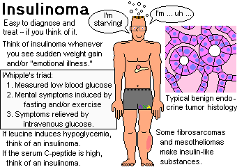
Look for Whipple's triad:
Often, patients become massively obese (why?)
The etiology of these tumors is obscure; there's not even an important genetic syndrome.
Of beta-cell tumor patients, 70% have a solitary adenoma, while the rest have either hyperplasia of many islands (* "nesidioblastosis"), or a beta cell carcinoma (the spectrum to malignancy: Cancer 104: 264, 2005).
Locating the tumor prior to surgery isn't easy, as they often do not show well on imagings. The favorite technique at the NIH is to inject the various pancreatic arteries with calcium and see which increases insulin levels in the hepatic veins (J. Clin. Endo. Metab. 94: 1074, 2009.) Once you've operated, expect a good outcome; aggressive insulinomas are rare (Arch. Surg. 147: 261, 2012).
* "Leucine-induced hypoglycemia" often heralds an insulinoma.
* Future surgeons: If the plasma insulin isn't down eight minutes after you remove the tumor, you didn't get it all. Surgery 132: 937, 2002. Removal by radioembolization: Am. J. Med. Sci. 340: 414, 2010.
* NOTE: Nesidioblastosis is a rare, poorly-understood cause of hypoglycemia in infancy. The islands and/or periductal tissue is packed with immature islet cells. Adults can get it too (Arch. Surg. 129 329, 1994), nobody knows how.
* Speaking of nesidioblastosis... Following the newly-popular Roux-en-Y gastric bypass surgeries, patients typically develop some hyperplasia of the islets, relieving any type II diabetes that may be present and sometimes even resulting in hypoglycemia (once attributed to dumping syndrome). Exactly what gut hormones cause this, and how, is being worked out. See NEJM 353: 249, 2005. Maybe it's ghrelin (the stomach's appetite-stimulating hormone, which stays low after bypass...) Update Science 320: 438, 2008.
PITFALL: The insulin produced by insulinomas is often a bit abnormal, and a lab assay can miss it: J. Clin. Endo. Metab. 88: 1464, 2003; Lancet 363: 363, 2004.
* NOTE: Many mesotheliomas and retroperitoneal fibrosarcomas, and occasionally other tumors, produce an insulin-like activity (probably somatomedin, but it varies).
NOTE: Be alert for factitious hyperinsulinism! To differentiate sly insulin administration from a beta-cell tumor, start by measuring plasma C-protein. To rule out oral hypoglycemic drug administration, check serum drug levels.
Gastrinomas (Zollinger-Ellison Syndrome; "G-cell tumors", etc.; older pathologist's review Cancer 68: 1329, 1991.)
An especially troublesome syndrome of multiple bleeding ulcers and diarrhea. The majority of gastrinomas are low-grade malignancies. The only way to tell is whether it has metastasized.
High basal acid secretion plus a marked increased in serum gastrin levels in response to secretin administration strongly suggests gastrinoma (Am. J. Med. 80: 11, 1986.
Screening asymptomatic persons for elevated gastrin levels merely turns up lots of older people with achlorhydria (Br. J. Surg. 77: 1, 1990 -- why?)
* Actually, a majority of gastrinomas, Zollinger-Ellison and otherwise, arise in the first or second portion of the duodenum.
Removal of all the tumor, if possible, is curative. Otherwise, these patients used to require total gastrectomy, but now they respond well to H2-blockers. Somatostatin analogues (notably "Octreotide", available from your pharmacist) are an effective alternative.
* Excess gastrin is an environment that encourages the growth of gastric carcinoids.
{09279} gastrinoma (to prove it, we'd need to
do an immunoperoxidase stain that stains gastrin brown and everything
else white)
{09282} gastrinoma
Glucagonomas ("alpha-two cell tumors"):
These produce mild diabetes, sore tongue, and necrolytic migratory erythema (death of the layer of cells 3/4 of the way up the epidermis). Don't miss this diagnosis (and people do: West. J. Med. 144: 746, 1986) -- even when metastatic, it responds well to somatostatin and its analogues (Acta Med. Scand. 218: 245, 1985; NEJM 314: 1686, 1986; Ann. Int. Med. 110: 35, 1989), and to oral zinc therapy. Update Am. J. Med. Sci. 321: 306, 2001; Gastroenterology 116: 1286, 1999. Two-decade survival despite liver metastases: Am. J. Med. Sci. 334: 225, 2007.
![]() Glucagonoma
Glucagonoma
Pittsburgh Pathology Cases
Multiple Endocrine Neoplasia Syndromes ("MEN", formerly "MEA", adenomas): some or all of the following in same family
Wermer's MEN I: pituitary adenoma, parathyroid adenoma, pancreatic endocrine tumors ( most often gastrinoma / Zollinger-Ellison; less often insulinoma, others)
Sipple's MEN II(a): Parathyroid adenoma, pheochromocytoma, medullary carcinoma of the thyroid
MEN IIb/III: Medullary carcinoma of the thyroid, pheochromocytoma, mucosal neuromas, Marfanoid habitus
More about these later....
Rarities:
Delta cell tumors ("somatostatinomas"): diabetes, diarrhea, gallstones, etc. Review: J. Surg. Onc. 43: 259, 1990.
Tumors secreting vasoactive intestinal polypeptide ("VIPomas"; "Verner-Morrison syndrome"): pancreatic cholera (horrible diarrhea), loss of potassium, achlorhydria -- excellent response to somatostatins. Update South. Med. J. 102: 761, 2009).
* PP-omas ("P-cell tumors"): no syndrome despite huge amounts of pancreatic polypeptide, an obscure hormone (Cancer 57: 129, 1986). Atropine suppression test: NEJM: 315: 287, 1986).
* Carcinoids: Cancer 104: 1180, 2005.
* Non-functioning tumors: If under 2 cm, think about leaving them alone (Surgery 150: 75, 2011), though the tendency now is to remove anything found on scan (Arch. Surg. 146: 534, 2011). More than a few turn out to be intrapancreatic accessory spleens (Arch. Path. Lab. Med. 134: 1474, 2010).
![]()
Even if you open up a practice
On Harley Street
No patient will come in with complaints
about his pancreas:
"I think it's my pancreas, Doc!"
-- unless he's a fellow professional
also educated
out of his natural mind; few patients
will be alarmed by the word -- how unlike
"the heart"
a word that means "the biscuit"
to the best of us.
Years from now
when you trundle in
thin and yellow, depressed,
for abdominal films,
you too will have forgotten
your pancreas; and the news "It's cancer
of the pancreas" will hit
like an old family secret you knew all along;
"I'm sorry, but it's cancer
of the sweetbread!"
"Not the sweetbread!" -- "Yes,
and, with proper medical management
early surgery
and a very rigid diet
you can look forward to at least
another three months"; when the pancreas goes
it goes.
Those among us who are diabetic
whom the pancreas torments
by degrees
cannot describe that Familiar; even a poet
is at a loss for a metaphor;
nothing short of a surgical exploration
will unearth
the thick spongy worm
buried deep in the viscera
silent behind its curtain of peritoneum;
-- with a head, a body,
and a tail,
using the man's face.
--Ron Charach, M.D.
NEJM 301: 508 (1979)
* SLICE OF LIFE REVIEW
08093 islet, normal
11748 pancreas, normal
14886 pancreas, normal
14886 pancreas, normal
14887 pancreas, normal
14888 pancreas, duct & islets
14889 pancreas, duct & islets
14890 pancreas, serous acini & islet
14891 pancreas, serous acini & islet
14892 pancreas, exocrine and endocrine
14893 zymogen cells, normal pancreas
14894 islet of Langerhans
14895 islet of Langerhans
14896 pancreas, zymogen granules
14897 pancreas, zymogen granules
14898 pancreas, central acinar cells
14899 pancreas, central acinar cells
15274 pancreas, normal
15275 pancreas, normal
15276 islet of Langerhans, #65
15277 pancreas, intercalated duct
15278 pancreas, centroacinar cell and duct
15279 islet of Langerhans, #65
15280 ?pancreas, ct in duct??
15793 pancreas, normal
20879 pancreas
20880 intercalated duct, pancreas
20881 acinar cell, pancreas
20882 islets of Langerhans, pancreas
20883 pancreas, centroacinar cell
20884 pancreas
20885 islet of Langerhans
20886 pancreas, interlobar duct
20887 pancreas, interlobar duct
25019 pancreas, normal
25020 pancreas, normal
25021 pancreas, normal
25794 columnar cells, pancreatic epithelium - norma
25973 pancreas, normal
25988 pancreas, normal
25991 acinar cells, normal pancreas
BIBLIOGRAPHY / FURTHER READING
I urge anyone interested in learning more about pathology of the pancreas to consult these standard textbooks.
In my notes, the most helpful current journal references are embedded in the text. Students using these during lecture strongly prefer this. And because the site is constantly being updated, numbered endnotes would be unmanageable. What's available online, and for whom, is always changing. Most public libraries will be happy to help you get an article that you need. Good luck on your own searches, and again, if there is any way in which I can help you, please contact me at scalpel_blade@yahoo.com. No texting or chat messages, please. Ordinary e-mails are welcome. Health and friendship!


| New visitors to www.pathguy.com reset Jan. 30, 2005: |
Ed says, "This world would be a sorry place if people like me who call ourselves Christians didn't try to act as good as other good people ." Prayer Request
| If you have a Second Life account, please visit my teammates and me at the Medical Examiner's office. |
Teaching Pathology
 Ed's Pathology Review for USMLE I
![]()
![]()
 | Pathological Chess |
 |
Taser Video 83.4 MB 7:26 min |
 |
Click here to
see the author prove you can have fun skydiving without being world-class. Click here to see the author's friend, Dr. Ken Savage, do it right. |


 Liver / Pancreas