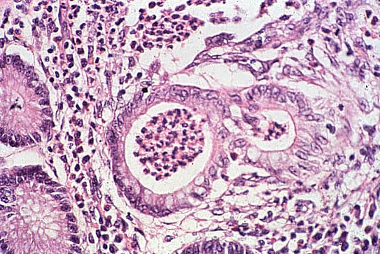
Crypt Abscess
It is very easy to see the central crypt. The tubular crypt of Leiberkuhn rounds the corner, in and out of the plane of section twice, giving it a double look.
Contrast the healthy crypts at the left edge with the abscessed crypt, and the neutrophils in the crypt abscess with the lymphocytes and plasma cells between the crypts.
Crypt abscesses are typical of ulcerative colitis, though not diagonstic. Several adjacent abscessed crypts precede loss of a patch of mucosa, forming the ulcers. This has not happened here yet, but there is a great deal of edema among these crypts. You can see this as spreading of the collagen fibers, especially toward the bottom.
| Visitors to Tom Demark's website since Sept. 1999 |