Betty, a 67 year old woman. Over the past year, increasing dyspnea on exertion, so that she cannot climb more than one flight of stairs. She has also lost 20 kg, and had paroxysmal nocturnal dyspnea. Six months ago, she fainted once. Four months ago, a local physician placed her on digoxin and a thiazide, without benefit. On admission, temp 36.7o, pulse 100, resp 24, BP 130/70. She is dyspneic on talking, and is thin. Jugular veins are distended to the mandible as she sits up. Coarse inspiratory crackles in the lower 2/3 of the lung fields. S1 normal, S2 normally split with a soft aortic valve component, soft S3, loud S4, grade 4 murmur of aortic valve stenosis. Liver edge 8 cm below the costal arch. Spleen barely palpable. EKG normal sinus rhythm, rate 80, PR interval 0.23 sec, QRS interval 0.09 sec, axis +50o, low voltage in extremity leads, inverted T waves in I, II, aVL, aVF, V3-V6. Cardiac catheterization data from day 4:
MGH 7-1972
What determines the voltage that is recorded on the limb leads? Mention several factors.
What sorts of things lengthen the PR interval? Can you suggest how they might work?
How do the "cath" values compare to normal?
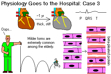
What effect does lack of compliance of heart muscle have on cardiac output?
What effect might loss of right atrial compliance have on production of atrial natriuretic peptide-factor-hormone?
If we had used this heart to test "Starling's Law", what surprises might we have had?
This lady's cardiac index is not so bad, yet she is in congestive heart failure. Can you suggest why? HINT: Think about the previous question.
Why do people with failing left ventricles get short of breath when lying down? Why might the shortness of breath increase so suddenly?
"Crackles" (once "rales") mean fluid in alveoli, hence bubbles popping. Explain its presence here.
The same process that made the heart muscle inflexible also rendered the arterioles and venules relatively unable to constrict or dilate. What effects would this have had?
Since "aortic stenosis" and "aortic insufficiency" and so forth refer to the aortic valve instead of the aorta itself, they are confusingly named. Do you know what we call a birth defect in which a short segment of the aorta is very narrow?
Main "Physiology Goes to the Hospital" Page
Case 1
Case 2
Case 4
Case 5
Ed's Pathology Notes
Ed's Home Page
Bryan's Home Page