Renoir, "In the Roses"
Renoir, "In the Roses"
Case: A 56-year-old woman, Mrs. Rose, visits you, her friendly neighborhood family doc, for her annual physical exam. The patient describes no health problems, but has noticed a little change in skin tone on the left side of her face over the last month or two. Mrs. Rose attributes that to getting a little older and spending too much time in the sun gardening. Social history: denies tobacco use and occasionally has a few glasses of wine, with her husband and friends. Surgical history her second child was delivered by C-section with no significant complications. Your physical exam reveals a slight lump on the left side of her face, which is fixed, but not painful to palpation. Careful examination reveals a slight sagging of the skin. The rest of the exam does not show any significant abnormalities. Differential diagnosis is enlarged lymph node versus a soft tissue tumor.
Test:
CT shows poorly enhancing area representing cystic change in the area of the parotid gland.
You call your local surgeon Dr. Ready Cut, who schedules her for surgery.
Pathology receives a total parotidectomy composed of a lobulated pink-tan specimen, including portions of the facial nerve.
Histologic sections reveal a tumor composed of two cell types’ myoepithelial cells and ductal cells in a cribriform pattern.
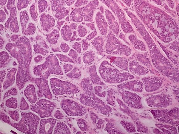
Immunohistochemical stains: markers for the characteristically dark, small-angulated myoepithelial cells as seen on H&E include myoepithelial markers, such as SMA, CD10. The epithelial component usually marks with keratins such as AE1/3, 1, 5, 7,8,10, 14, 18 and 19.
In tumors that undergo a high-grade transformation, with complete loss of myoepithelial cells are often p53 positive. Genetic testing of the mass shows t(6;9) (q21-24;p13-23).
Treatment: Surgical resection with radiation therapy. Resection showed a light-tan, solid, well-circumscribed but unencapsulated mass.

Alexander Palmer KCUMB '15