WHITE CELL DISORDERS I & II
Title: White Cell Disorders I & II
Date & Time: Monday, November 12, 2012 at 12 nooon (White Cell Disorers I)
Date & Time: Wednesday, November 14, 2012 at 12 nooon (White Cell Disorers II)
Lecturer: The Pathology Team
QUIZBANK -- Blood & Lymph #'s 133-139, 178-333
INTRODUCTION
You will refer to this material every time you feel a large lymph node or spleen, or have a patient with
an abnormal CBC.
"Leukemias and lymphomas" is the most difficult unit in Medical Pathology except for glomerular disease. You can't learn
it if you are not continually asking yourself, "Why?"
You are already familiar with the development of the different kinds of white cells, and the locations
of lymphoid tissue throughout the body (lymph nodes, Waldeyer's ring, Peyer's patches, spleen, large
airways).
T-cell zones: thymus, lymph node parafollicular cortex, splenic white pulp near arteriole
B-cell zones: germinal centers and their mantles, splenic white pulp at its margins
Among circulating lymphocytes, 80% are T-cells, and 20% are B-cells.
* You are also familiar with the common reaction patterns of various white blood cells: acute
inflammation, pus, granulomas, and accumulations within phagocytes. (There's no need, for example,
to talk right now about xanthomas, lipogranulomas, etc., etc.)
 Mycobacterial lymphadenitis
Pittsburgh Pathology Cases
In discussing diseases that affect numbers of white blood cells in the peripheral blood, it is much more
useful to talk about ABSOLUTE CELL COUNTS than "percentage counts".
Of course, you can estimate the absolute count by multiplying the total WBC count x the % for a
particular cell.
Healthy absolute counts:
Basophils: * few- 100/cu mcL (heads up -- basophil granules are soluble and can wash out during slide preparation)
Eosinophils: few- maybe 400 (fewer in AM, more in PM)
(* "Hypereosinophilia"
was once defined to be more than 1500 for more than six months without an obvious reason,
and some evidence of organ involvement; today, as soon as one of the
hypereosinophilic syndromes causing organ damage is suspected,
diagnose if you can and start treatment even if you can't)
Lymphocytes: 1200-3400 (* 3000-7000 for kids)
Monocytes: 100- 590
Neutrophils: 1800-6500
Note that "95% lymphocytes" might mean either agranulocytosis (if the total white count is 2000) or
chronic lymphocytic leukemia (if the total white count is 100,000). This is why I like
white cell differential counts reported in absolute numbers, and why all labs do this nowadays.
* Current smokers average 25% higher neutrophil counts; those who've quit in the last five years still
average higher (Am. J. Clin. Path. 107: 64, 1997). This won't matter in your clinical decision-making.
A good "normal range" for total white count is 4000-11000/cu mcL. "Leukocytosis" is present when the
white count exceeds 12,000/cu mcL.
The most important "white cell diseases" are neoplastic. These are:
(1) the MALIGNANT LYMPHOMAS (HODGKIN'S AND NON-HODGKIN'S), solid tumors of lymphocytes (the rare
tumors that truly arise from monocyte-macrophages are also included here; no one knows the true cell of origin of
the malignant cells of Hodgkin's disease, which is also included here)
(2) the LEUKEMIAS and their close relatives, the MYELOPROLIFERATIVE
DISORDERS, in which sick hematopoietic
stem cells proliferate
(3) the PLASMA CELL DISORDERS, which typically produce antibodies and/or fragments thereof
(4) the LANGERHANS CELL HISTIOCYTOSIS FAMILY ("histiocytosis X"; "disseminated histiocytosis") of quasi-cancers, much
less common than the others
Probably because it is so easy to harvest the cells,
and since chemotherapy has been more successful for these
diseases than for most other cancers, a tremendous amount
of study has gone into clarifying their molecular pathology.
There can be no such thing as a truly benign neoplasm of white blood cells, since by their very
nature they infiltrate tissues. Some of these entities (for example, the acute leukemias)
are far more aggressive than others ("benign plasmacytoma",
"benign monoclonal gammopathy").
White cell markers oversimplified:
{16282} E-rosette, around a T-cell
CD3, CD4, CD8, αTcR, βTcR, γTcR, δTCR, others: the more mature T-cells (various kinds)
CD1a (T6):
some T-cells, all Langerhans macrophages
CD5: mantle cell lymphoma, many CLL
* CD10 (CALLA): most B-cells (mantle cell lymphoma is negative)
CD15: Most Reed-Sternberg cells; some others
* CD19: B-cells, but not plasma cells
* CD20: all but the most primitive B-cells, but not plasma cells
* CD22: most B-cells (EBV receptor)
* CD34 : primitive blood cells -- great for counting "blasts" in leukemia / preleukemia
CD45 ("common leukocyte antigen" / LCA): all white cells (* exception: Reed Sternberg cells and some leukemias)
* CD65: Most consistent marker for the natural-killer lymphocytes, non-B, non-T cells
making up maybe 10% percent of your circulating white cells. (They tend to be big and have granules.
Update on their neoplasms: Cancer 112: 1425, 2008).
CD68: common macrophages
* CD79a: The mantle lights up best
BCL-2 (apoptosis-preventer): turned OFF during hypermutation (i.e., germinal centers); turned ON in most nodular lymphomas
surface Ig(M, etc): B-cells
kappa, lambda: mature B-cells, plasma cells -- especially useful for showing clonality (i.e., neoplasia)
* Future pathologists only: To look for clonality,
you can also have the lab check to see if the
rearrangements
that produce the specificity of a lymphocyte for a
specific antigen are clonal
(IgH Gene Clonality for B-cells, TcR-gamma Gene Clonality
for T-cells).
cyclin D1: mantle cell lymphoma stains strongly
cytoplasmic Ig: plasma cells
* nonspecific esterase: monocytes
Fc receptor: B-cells, monocytes
TRAP: hairy-cell leukemia
HLA-D/DR /Ia: Langerhans cells and other antigen-presenting macrophages; some other cells
lysozyme: monocytes
* alpha1-antichymotrypsin: monocytes
erythrophagocytosis: monocytes
(myelo-)peroxidase: granulocytes
* Sudan black:
granulocytes
* chloroacetate esterase: neutrophils, basophils, mast cells
platelet markers: megakaryocytes
* PAS+ diffusely: erythrocytes, megakaryocytes/platelets
* PAS+ chunks ("blocks"): immature lymphocytes or M6 leukemia
S-100, CD1/T6: dendritic ("Langerhans") macrophages
I would ask you NOT to worry about differentiation markers
beyond what's been listed above. A pathologist MUST know them, as a Hodgkin or non-Hodgkin lymphoma cannot
be properly classified without immunohistochemistry (update Arch. Path. Lab. Med. 132: 441, 2008).
A sub-subclassification for epidemiologists: Blood 110: 685, 2007.
* There has been talk of diagnosing and classifying lymphomas based on little biopsies
(less invasive than taking out a whole lymph node.) As you'd expect, unless there's an easy trademark
finding or two (mantle-cell lymphoma, T-lymphoblastic lymphoma), it can't be done reliably
(Am. J. Clin. Path. 128: 474, 2007).
* {16517} neutrophil, chloroacetate esterase stain
 Neutrophilia
Text and photomicrographs. Nice.
Human Pathology Digital Image Gallery
NEUTROPENIA: A low absolute neutrophil count in the peripheral blood for any reason. (NOTE:
"Leukopenia" is a not-very-useful word that describes any low total white count.)
Possible causes include
SUPPRESSION OF GRANULOPOIESIS
"The aplastic anemias" (better, "bone marrow failure")
Bad stuff in the marrow
Space-occupying lesions ("myelophthisic anemias")
Hematologic malignancies that suppress granulopoiesis (i.e., some leukemias and lymphomas)
DNA problems
Cancer chemotherapy
Radiation sickness
"The megaloblastic anemias"
Hereditary cyclic (q. 3 wk., severe; dominant mutation usually in the ELA2 elastase gene, molecular biology Blood 92: 2629, 1998; one cause is mutated neutrophil elastase that itself damages the cellular machinery; also Nat.
Genet. 35: 90, 2003; Blood 108: 493, 2006)
* Shwachman-Diamond (genetic, also fatty pancreas)
Typhoid fever
Occasional virus infections (mild suppression, especially parvo B19 (Am. Fam. Phys. 75: 373, 2007))
* Some childhood acute leukemias going back to the stem cells
* MYELOKATHEXIS (group of genetic diseases with accelerated neutrophil precursor
apoptosis; surviving neutrophils are hypersegmented and have very long bars between nuclear lobes:
Blood 95: 320, 2000; Am. J. Hem. 62: 106, 1999).
* Kostmann's -- genetic disease (several loci); almost all of the developing neutrophils die at the myelocyte stage.
The usual cause of very low absolute neutrophil count at birth and after.
Idiopathic
* The lab machine didn't count them.
- The blood sat in EDTA too long and the neutrophils stuck together.
- The patient has one of leukemias / preleukemias in which the neutrophils
don't make much myeloperoxidase, and the machine used the myeloperoxidase reaction
to count neutrophils
EXCESS DESTRUCTION OF NEUTROPHILS
Autoimmune (rare, think of lupus)
Hypersplenism (see below)
Sequestration in a rapidly-growing abscess (??)
Idiopathic
DRUGS: The mechanisms are typically obscure (Ann. Int. Med. 146: 657, 2007).
PERSONAL-TRIVIAL
AGRANULOCYTOSIS is a time-honored misnomer for neutropenia sufficiently severe to put a person at risk
for serious infection (i.e., neutrophil counts of 1000 or less, often much less; <500 is a big emergency).
The first sign is typically mouth ulcers ("there's lots of germs in there") with their pseudomembranes
laden with infectious bacteria and/or fungi.
* There may also be ulcers in the cecum; these can actually kill
("acute typhlitis") by providing a portal of entry to the blood for bacteria.
Later, the body is overwhelmed by bacteria, with death ensuing in a few days. Until the very end,
patients are likely to complain only of "just not feeling quite right".
The usual cause of "agranulocytosis" problems is medications. Future docs: If you notice that somebody has
an absolute neutrophil count <1800 or so, stop all medications that can be stopped,
and check again in a week.
LYMPHOCYTOPENIA is less common and less perplexing than neutropenia. Think of hereditary
immunodeficiency, HIV, radiation injury, marasmus/kwashiorkor, Cushing's syndrome, or just "stress".
Apart from AIDS, the most important cause clinically is "multiple organ failure"
of the severely sick / very septic; lymphocytes undergo apoptosis throughout
the body, and this is mirrored in lymphoid depletion at autopsy (J. Imm. 174:
3765 2005).
LEUKOCYTOSIS: It's worth remembering the following NON-NEOPLASTIC
CAUSES OF ELEVATED WHITE CELL COUNTS. Most of them make sense:
You remember that in health, about half the neutrophils in the blood are circulating, and the other half
are marginated, at any time.
LOTS OF NEUTROPHILS ("granulocytosis"):
- pyogenic bacterial infection (the usual cause)
- burns
- widespread tissue necrosis from any cause
- late pregnancy (common, mild)
- really bad "collagen-vascular disease"
- just plain "stress", nausea, and/or physical pain (un-marginates neutrophils)
glucocorticoids and epinephrine do the same thing;
glucocorticoids also prevent neutrophils from entering tissues
NOTE: Typhoid patients and some super-septic patients may become neutropenic because granulopoiesis
is suppressed and/or all the neutrophils have emigrated from the blood. Beware
of relying on white count as your chief marker for infection!
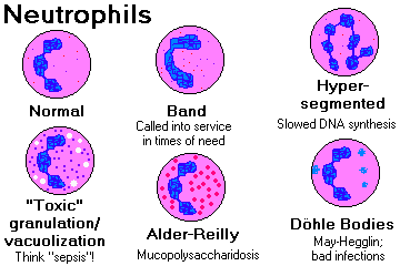
NOTE: The super-sick, septic patient is likely to have TOXIC
GRANULATION (extra-prominent azurophilic
granules), CYTOPLASMIC VACUOLES ("from doing all that phagocytosis"), and/or
DOHLE BODIES (rough
endoplasmic reticulum remnants). By contrast, if the neutrophil count
simply rises from acute pain and "stress", there will be
no toxic granulation, vacuolization, or left shift.
More about these in "Clinical Pathology".
{13646} Dohle body
{13661} Dohle body
{16213} Dohle body
* Future pathologists: The latter two "Dohle bodies" are fakes; they are
from cases of May-Hegglin's (say "Muh-HAY-lun") semi-disease, an
autosomal dominant trait with too-few, too-big platelets and lots of "Dohle bodies" and big granules; the neutrophils
function normally. May-Hegglin "Dohle bodies" are actually
non-muscle myosin A, gene mutated in May-Hegglin: Nat. Genet. 26:
106, 2000; Blood 97: 1147, 2001. There are several different phenotypes
at the locus (Blood 102: 529, 2003).
NOTE: LEFT SHIFT refers to presence of immature white cells ("bands") in the peripheral blood, i.e., they're being
mobilized early from the bone marrow. To tell an extreme case (WBC>up to 100,000 or so, i.e., a
LEUKEMOID REACTION, as in sepsis, overwhelming TB , or carcinomatosis) from chronic granulocytic
leukemia (see below), remember the following:
(1) In chronic granulocytic leukemia, the LEUKOCYTE ALKALINE
PHOSPHATASE tends to be low. In sepsis and
the non-leukemic myeloproliferative disorders, it tends to be high.
Leukocyte alkaline phosphatase is a completely different test from the "serum alkaline phosphatase" on
the chemical profile. DON'T talk about them together.
(2) In chronic granulocytic leukemia, the ABSOLUTE
BASOPHIL COUNT is generally high, too. This would
be unusual in sepsis.
(3) In chronic granulocytic leukemia, there is virtually always a switch of material between
chromosomes 9 and 22 (i.e., the PHILADELPHIA CHROMOSOME (Ph')
or at least its molecular equivalent). You
won't see this except in cancer.
(4) And of course, toxic granulation (very easy-to-see granules on stained blood; nobody really
knows why)/ toxic vacuolization
says "infection", not "leukemia".
(5) When in doubt, it's a leukemoid reaction. An indolent leukemia can wait for a few days; deadly infection can't.
Philologists: RIGHT SHIFT refers to the hypersegmented granulocyte nuclei of pernicious anemia (etc., any
major impediment to normal DNA synthesis will produce this "megaloblastic" change). "Right" and
"left" derive from spaces on the old do-it-by-hand tally sheets.
The machine-counting of immature neutrophils has always been
a challenge. Review and new equipment: Am. J. Clin. Path. 128:
454, 2007. Today we know that the band count is very low in health, around one white cell in 500.
LOTS OF EOSINOPHILS (big review Mayo Clin. Proc. 80: 75, 2005):
The "Loeffler" family of eosinophil-mediated diseases -- now being sorted out;
while most remain idiopathic, a few have known mutations
CHRONIC EOSINOPHILIC LEUKEMIA, an entity removed from the Loeffler's
wastebasket by the discovery of FIP1L1-PDGFRalpha (Haematol 95:
696, 2010) -- treat with imatinib.
type I immune injury
food allergy, hay fever, eczema, extrinsic asthma (supposedly -- you won't be impressed)
bronchocentric granulomatosis (aspergillus superinfection in asthma; this one's important)
* hyper-IgE ("Job's") immunodeficiency
Tissue parasites
ascariasis
filariasis (includes "tropical eosinophilia" of the Far East -- future pathologists: filaria worms will be pushed to the "feather edge" of the smear)
onchocerciasis
strongyloidiasis
trichinosis
echinococcus
visceral larva migrans (dog and cat roundworms)
cutaneous larva migrans (dog and cat hookworms)
Drug allergy (most any; but notoriously gold therapy for arthritis, where eosinophilia is almost expected)
Hodgkin's disease (a large minority of cases)
Churg-Strauss (a vasculitis, often with granulomas, usually with ANCA;
it's not clear whether this is a separate disease, or simply the way
Wegener's / polyarteritis manifests in folks with allergies)
Dermatitis herpetiformis
Familial hypereosinophilia (locus unknown, autosomal dominant, mild: Blood 103: 4050, 2004)
* Well's eosinophilic cellulitis
Eosinophilia-myalgia syndrome (from the tainted tryptophan)
* Any AIDS patient with a rash (Am. J. Med. 102: 449, 1997)
* Pemphigus (I don't know why)
* Dermatitis herpetiformis
* Crohn's
* Acute liver transplant rejection (almost all have it, no one knows why)
Dermatomyositis
Polyarteritis nodosa (don't miss this one)
* Kimura's angiolymphoid hyperplasia with eosinophilia (very high IgE, eosinophil-lymphoid pseudotumors of head and neck,
germinal centers loaded with eosinophils;
marked peripheral eosinophilia;
common in middle-aged Asian men, Asia, rare elsewhere;
making the call Pediatrics 110: e-39, 2002; probably a low-grade
lymphoproliferative disorder Am. J. Surg. Path. 26: 1083, 2002; Arch.
Path. Lab. Med. 131: 650, 2007)
* mastocytosis with eosinophilia (molecular signature known, response to imatinib/Gleevic likely)
* T-cell neoplasms making interleukin-5: NEJM 341: 1141, 1999
* "Clonal eosinophilia" -- FIP1L1-PDGFRA fusion gene
* Eosinophilic leukemia -- looks like CML without proof of clonality
NOTE: In the developed world, among clinically healthy patients with isolated elevated
eosinophil counts, you will often not find the cause.
NOTE: I did CBC's for years on medical students, many of whom have hay fever, etc., and
have never found one with an elevated eosinophil count.
NOTE: Remember that eosinophilic counts are up in the afternoon and down in the
morning because the morning's cortisol surge suppresses them;
I'd suggest taking a serious look at an absolute eosinophil count over 350 or so in the morning,
and over 650 in the afternoon.
NOTE: The "Loeffler's eosinophilic" problems are a curious, mixed-bag of diseases with excessive
numbers of eosinophils in various tissues that cause tissue damage.
The most common form
is probably caused by a benign neoplasm of some sort,
hidden somewhere,
with a fusion gene called FIP1L1-PDFGRA (Cancer 110: 955, 2007)
This has sometimes responded well to imatinib.
Sometimes the underlying problem is a proliferation of
mutated T-cells producing excessive eosinophil attractants (NEJM 330: 35, 1994).
* In other cases, the eosinophils themselves seem to be the
mutated clone: Blood 93: 1651, 1999.
In 2004, I predicted the success of
the anti-IL5 antibody mepolizumab as treatment (J.
Allerg. Clin. Imm. 113: 115, 2004); it has been a
spectacular success (NEJM 358: 1215, 2008).
{14099} eosinophilic leukocytes (buffy coat)
{09207} eosinophil granule with crystal (electron micrographs; these crystals will combine to form large
Charcot-Leyden crystals under some conditions)
 Eosinophilia
Text and photomicrographs. Nice.
Human Pathology Digital Image Gallery
LOTS OF MONOCYTES:
typhoid fever
bad granulomatous problems
* chronic autoimmune disease
* rickettsial disease (often; red flag)
* disseminated cancer (occasionally)
LOTS OF LYMPHOCYTES:
"infectious mononucleosis" (see below)
whooping cough ("pertussis"; little cleaved lymphocytes; the toxin keeps the T-cells from homing to lymphoid tissue; Am. J. Clin. Path. 114: 35, 2000)
infectious lymphocytosis (mild kids' disease, with T-cells,
caused by various non-herpes viruses notably coxsackie B2 ; a "chronic form" also exists without
marrow abnormalities; leave this to the pediatric hematologists; Acta. Paed. 74: 633, 2008.)
"transient stress lymphocytosis" (absolute counts 4000-10000; on the evidence
we've overlooked this for years; all major lymphocyte subsets go up,
and neutrophils go up too: Am. J. Clin. Path. 117: 819, 2002)
* really bad "collagen-vascular disease"
* phenytoin ("Dilantin") or para-amino salicylic acid ("PAS") therapy
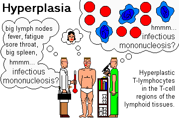
NOTE: INFECTIOUS MONONUCLEOSIS is a family of diseases featuring fever, malaise, fatigue,
lymphadenopathy, and circulating benign atypical lymphocytes. The syndrome results from first
meeting one of these four micro-organisms: (1) Epstein Barr virus ;
(2) cytomegalovirus
 ;
(3) toxoplasmosis
 ;
(4) HIV.
BENIGN ATYPICAL LYMPHOCYTES are activated cells (B- or T-) seen typically
in the blood in "infectious mononucleosis" and certain other infections;
you may see a few in any viral illness.
There is no such thing as a "typical atypical lymphocyte." There are three "Downey" types:
- Lymphocytes just a bit larger than usual, with a nuclear cleft and dark cytoplasm
- Most familiar: the cytolasm is abundant and pale,
bluer where the red cells indent them;
the nucleoli are small, the nucleoplasm is reticulated
- immunoblasts -- big cells, bit nucleoli, plenty of blue-staining cytoplasm
Probably more important in spotting infectious mono and telling it from leukemia
when you're a beginner is that when there are bunch of "atypical lymphocytes",
no two look the same.
LOTS OF BASOPHILS:
chronic myelogenous leukemia
other "chronic myeloproliferative disorders"
* supposedly in lots of other things; this will not be important clinically.
NOTE: None of these "classic findings" is either particularly sensitive, or particularly specific, for any
particular disease. Use this information in the setting of the "whole person".
ODD NEUTROPHILS:
We have already mentioned CHRONIC GRANULOMATOUS DISEASE, a poorly-named
group of defects in the ability of neutrophils to kill common bacteria,
with the macrophages needing to become involved as well.
The most familiar is "X-linked chronic granulomatous disease",
which has now been cured by gene therapy (Nat. Med. 12: 401, 2006).
|
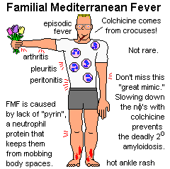
FAMILIAL MEDITERRANEAN FEVER, long-mysterious, has now
yielded up its secrets.
The cause is a lack of pyrin, a neutrophil
protein that slows down neutrophils
when enough have reached an area. Gene found Cell 90: 797, 1997;
molecular genetic diagnosis: Ann. Int. Med. 129: 539, 1998.
Lacking pyrin, neutrophils mob body cavities every once in a while.
In addition to fever, patients may have pleuritis, arthritis,
peritonitis, and/or a hot rash (looks like a strep infection) on the ankles.
Colchicine, famous for its ability to slow down neutrophils
(as in acute gout), controls the attacks and prevents the dread
complication of secondary amyloidosis.
As you can imagine, acute FMF can mimic many diseases. The amyloidosis AA
that often develops in these patients can mimic most of the rest. Don't miss it.
* A similar, thankfully-rare periodic fever syndrome
is caused by a mutation in the TNF-receptor (TNFR1): Blood 108:
1320, 2006. Yet another is caused by mutated cryopyrin
(includes serious neurologic problems; anti-interleukin-1 treatment
brings about full resolution: Neurology 74: 1267, 2010). | |
|
You recall CHEDIAK-HIGASHI SYNDROME, in which there are several problems with
organelle membrane synthesis.
synthesis.
Neutrophil lysosomes are large and prominent (they fuse with each other) and do not fuse with phagosomes,
so there's poor bacterial killing and a lot of infections.
Melanosomes don't form properly, so there is partial albinism.
The lack of platelet dense granules results in a bleeding tendency.
* The gene has been cloned (LYST, lysosomal traffic regulator). Most of these patients go on to
develop a lethal non-neoplastic hyperplasia of the lymphocytes.
Marrow / stem cell transplant is now routine and prevents this.
Review Blood 95: 979, 2000.
* There is a report that long-term survivors of bone marrow transplantation
develop a neurodegenerative disease after decades (Blood 106:
40, 2005). Stay tuned.
In the autosomal dominant PELGER-HUET ANOMALY, the neutrophil nuclei fail to segment normally, producing
"peanuts" and "pince-nez eyeglasses". ("Look! This clinically healthy patient has a horrible left shift / leukemia!")
This is a fairly common laboratory curiosity (maybe one person in 5000), and of no significance.
Explain to the physician and the patient, and check their close kin before somebody gets sick
and everybody gets confused (Am. J. Clin. Path. 137: 358, 2012). (* Double doses get no
segmentation whatever. And they don't seem to have any obvious troubles
with bacteria or
anything else: Acta. Hem. 66: 59, 1981. "Look! This clinically healthy patient has all myelocytes!"
"No, look around, they're pelgeroid.")
* Acquired / pseudo-Pelger-Huet can been seen when there
are mutations (i.e., myelodysplasia, leukemia) or for some mysterious
reason as a medication side-effect (Arch. Path. Lab. Med. 130: 93, 2006;
Am. J. Clin. Path. 135: 291, 2011). These tend to be a minority of neutrophils,
tend to be hypogranular, and tend to have denser chromatin. Let us worry about them.
| | 
|
{16208} Pelger-Huet, one dose
{16209} Pelger-Huet, one dose
{13658} Pelger-Huet, two doses
* ALDER-REILLY ANOMALY merely refers to large, mucopolysaccharide-laden granules in some
of the storage diseases (Hunter's, Hurler's, Tay-Sach's, occasionally as an acquired
trait in myelodysplasia). You will see it in all five types of white blood cells.
Don't mistake this for "toxic granulation."
* Thankfully rare: Lack of endothelial adhesion molecules for
phagocytes (J. Clin. Invest. 103: 97, 1999) or lack of
CD18 integrin on neutrophils (Blood 91: 1520, 1998).
Bacilli in neutrophil vacuoles: Usually DF2 (dog bite)
* And you know that drumsticks are the inactivated X-chromosomes of lyonization.
MORULAE OF EHRLICHIOSIS can help you diagnose
this famous "spotless fever"; this "granulocytic" variant
of ehrlichiosis can be fatal (NEJM 334: 209, 1996). "Morule" is Latin for "mulberry".
 Ehrlichiosis
Morules in macrophages
CDC photo
NORMAL LYMPH NODE ANATOMY
LYMPH NODES are soft (i.e., reticulin-framework) ovoids, up to about 2 cm in health. Afferent
lymphatics penetrate and travel within their capsules (metastatic cancer first sets up here). Afterwards,
lymph percolates through the cortex, and then the medulla, leaving by the hilum.
Within the cortex, there are generally some germinal centers ("lymphoid follicles"), sites of actively-proliferating B-cells.
Each germinal center is surrounded by a mantle of resting B-cells, which are in
turn surrounded by "parafollicular" T-cells. (If there is no antigenic stimulus, you'll see only "primary
follicles" of sleepy B-cells in the cortex.)
The next time you get to look at a germinal center under the microscope, check out those proliferating
B-cells. The sequence from small B-cell to plasma cell is interesting and unsung in most histology
courses. You'll need to know this to understand classical acconts of lymphomas:
Resting small B-lymphocyte
 Small cleaved ("clefted", i.e., folded-nucleus) B-lymphocyte
 Large cleaved B-lymphocyte
 Small non-cleaved B-lymphocyte
[NOTE: This cell is as large as a large cleaved B-lymphocyte]
 Large non-cleaved B-lymphocyte
 B-immunoblast


 Memory B-cells . . and . . Plasma cells
Within the medullary cords, expect to see a mix of B- and T-cells and plasma cells. The sinusoids are
lined by fixed phagocytes.
Despite the elegant pictures in histology books, lymph nodes are seldom "normal", especially in
adults.
LYMPHADENITIS: Inflammation of the lymph nodes
ACUTE LYMPHADENITIS described in "Big Robbins" is not much more than the hyperplasia in a reactive
node.
Localized lymphadenitis is most often due to a bacterial infection in the area drained by the lymph node.
Really bad cases have polys and even abscess formation within the nodes. The end result will be a
scarred-up lymph node. You have one or more.
Generalized lymphadenitis suggests a systemic viral infection.
"Mesenteric adenitis", often indistinguishable from acute appendicitis, is caused by Yersinia
enterocolitica.
Acute lymphadenitis, since it comes up suddenly and stretches the capsule, is likely to make the node
tender.
CHRONIC NON-SPECIFIC LYMPHADENITIS falls in one of three distinctive patterns.
FOLLICULAR HYPERPLASIA (i.e., lots and lots of big follicles) results from longstanding contact with
organisms or "other things" that stimulate the B-cells. If perplexed, think of:
- toxoplasmosis
 (* look for mini-granulomas touching the germinal centers
at their edges, this is supposedly pathognomonic; some of these are groups of
macrophages; some are big "monocytoid B-cells" especially in the medulla)
- rheumatoid arthritis (often lots of plasma cells)
- syphilis
 (plasma cells in the medulla, mini-granulomas, spirochetes)
- AIDS-related complex / persistent generalized lymphadenopathy of HIV infection.
- common variable immunodeficiency (ineffective B-cell activation)
{36371} toxoplasmosis; many bugs in a cell
{40654} toxoplasmosis; tissue reaction (lame-looking granulomas)
PARACORTICAL LYMPHOID HYPERPLASIA (i.e., lots and lots of lymphocytes, including turned-on ones, in the
T-cell regions of the cortex -- often easiest to recognize the the presence
of prominent blood vessels) results from longstanding contact with organisms or "other things" that
stimulate the T-cells. If perplexed, think of
- infectious mononucleosis, CMV
- phenytoin ("Dilantin") exposure
- weird reactions to a vaccine
- infectious mononucleosis family (those angry T-killers, etc.; * expect to see lots of big lymphoid
cells with pale cytoplasm, plenty of necrosis and mitotic figures; see any
CMV
 cells?; this can be a real
fooler for lymphoma, see Arch. Path. Lab. Med. 117: 269, 1993)
- lupus (look for frank vasculitis and even regions of infarction)
SINUS HYPERPLASIA (formerly "sinus histiocytosis"; i.e., sinusoids with swollen endothelial cells and lots of histiocytes). If perplexed,
think of:
- nodes draining a cancer (by no means specific!)
- * nodes injected with permanent radiology contrast medium ("lymphangiogram dye") or for some reason containing mineral oil ("oleogranulomas")
- Whipple's disease (Whipple cells)
- * Castleman's giant angiofollicular lymphoid hyperplasia (far beyond your learning objectives; NEJM
330: 642, 1994); one cause (especially when multifocal and rich in plasma cells)
seems to be herpes 8
 / KSHV (Am. J.
Path. 151: 1517, 1997; West. J. Med. 167: 38, 1997;
Blood 93: 3643, 1999). It is a famous cause of paraneoplastic
pemphigus (Lancet 363: 525, 2004).
 Castlemanosis
Great photos
Pittsburgh Pathology Cases
- * hemolysis (Coombs-positive, or macrophages rendered hungry by infection; the latter situation
is "erythrophagocytic reticulosis")
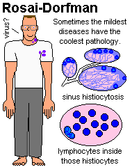
- * "sinus histiocytosis with massive lymphadenopathy and lymphocyte emperipolesis"
(Rosai-Dorfman disease): a presumably
viral, generally self-limited semi-disease of young people. B- and T-cells live inside
macrophages ("emperipolesis"), which in turn light up with S100 and for lipid. There is a more
ominous
extranodal form as well. No one understands it.
| |
|
 Rosai-Dorfman
S100 for dendritic macrophages
Wikimedia Commons
MIXTURES OF THE ABOVE cause diagnostic problems. In all the above, capillary endothelial cells are likely
to be hyperplastic (rare in cancer).
The most common cause of "unexplained" lymph node
enlargement, especially in the groin: DERMATOPATHIC LYMPHADENITIS, melanin and sebum-laden nodes
draining chronically inflamed skin. You're likely to see a mix of reaction types.
{35609} dermatopathic lymphadenitis (the red-brown is melanin, the white is sebum)
WARNING: Any of these patterns can be (and occasionally is) mistaken for malignant lymphoma by
the inept. Note that the finding of mitotic figures or necrosis doesn't necessarily point to malignancy,
while the presence of a variety of cell shapes actually suggests a benign diagnosis. Know your
pathologist, and ask for consultation if you are in doubt.
GRANULOMAS
Granulomas with central CASEOUS NECROSIS are probably tuberculosis, some other mycobacterial
infection (atypical mycobacteria, leprosy)
Well-made granulomas with NOTHING else are probably sarcoidosis. Also remember Crohn's, berylliosis,
and nodes draining Hodgkin's disease.
Granulomas with PUS in their centers are probably caused by one of the
following: (1) lymphogranuloma
venereum ,
(2) cat scratch fever, (3) brucellosis,
(4) plague
 ,
(5) tularemia
 ,
(6) glanders-melioidosis, and
(7) other yersinia infections. If you can find none of these, consider (8) X-linked chronic
granulomatous disease (the neutrophil dysfunction problem).
* Granulomas with central necrosis with much karyorrhexis but no pus:
Kikuchi-Fujimoto. See below.
You have probably already seen the Warthin-Finkeldey giant cells
of measles within germinal centers. They show variable immunologic markers --
B-cell, T-cell, and/or dendritic macrophages. Some have intranuclear measles virus inclusions; some do not.
* KIKUCHI-FUJIMOTO NECROTIZING HISTIOCYTIC LYMPHADENITIS (J. Am. Acad. Derm. 59:
130, 2008; Am. J. Clin. Path. 131: 174, 2009): Nobody knows the cause of what seems to
be a viral illness (the herpes family
exonerated Arch. Path. Lab. Med. 131: 604, 2007); the molecular
biology is not that of a lymphoma: Am. J. Clin. Path. 117 627, 2002.
Nepalese study: Arch. Path. Lab. Med. 127: 1345, 2003.
Big review Am. J. Clin. Path. 122: 141, 2004.
It looks like lupus in the lymph node, but there's lots more cytotoxic CD8+ than CD4+
T-cells.
I've got a story about this I'll tell you personally.

NON-HODGKIN'S LYMPHOMAS: By definition, monoclonal, malignant tumors of the B- or T-cells,
and not of plasma cells, and not Hodgkin's disease. By custom,
soft tumors of monocytes are included here because they look similar.
These are the common primary tumors arising in the lymphoid tissue
(lymph nodes, tonsils, adenoids, spleen, Peyer's patches, non-epitehlial thymus) and there are some
special cases. (We cover CNS lymphomas in the "neuro" section; the outlook for primary CNS
lymphoma is still "dismal": Cancer 110: 1803, 2007.)
There are about forty kinds at most recent count, each with its
own personality. Together, the non-Hodgkin's
lymphomas are common. Update, with a focus on molecular markers: Br. Med. J. 362: 139, 2003; also Lancet 362:
139, 2003; J. Clin. Path. 58: 561, 2005.
Most pathologists use the 2008 World Health Organization classification.
It is elaborate even by WHO standards and only the highlights can be covered here.
 Follicular lymphoma, spleen
AFIP
Wikimedia Commons
The non-Hodgkin's lymphomas are a subject of perennial fascination for pathologists. Making the
diagnosis ("benign or malignant?") is often tough, and classifying the non-Hodgkin's lymphomas
(hereinafter "lymphomas") was a major international competitive sport through the 1980's.
Today, the ongoing fascination is in the chromosomal translocations that
are the primary way in which white blood cells acquire mutations. In most
of the leukemias and lymphomas, the genome is usually NOT destabilized.
Today, several translocations actually define a particular leukemia or lymphoma.
* Now-classic review of the translocations: Arch. Path. Lab. Med. 127: 1148, 2003.
Molecular probes now routinely detect translocations missed by conventional cytogenetics
(Am. J. Clin. Path. 135: 921, 2011).
Students often find this subject especially difficult to understand. Hence, the focus in this section on
"Rules".
RULE: All monoclonal proliferations of lymphocytes are best considered malignant. (Some monoclonal
plasma cell proliferations might be benign.)
RULE: Most non-Hodgkin lymphomas are somewhat more common in men, with the most pronounced difference
probably being T-lymphoblastic lymphoma (> 2:1).
RULE: Blacks and children almost never get nodular lymphomas.
RULE: A few specific lymphomas have one or more special risk factors (i.e., helicobacter causes
the MALT lymphoma of the stomach;
gluten enteropathy "sprue" causes a curious pair of T-cell lymphomas: Gastroent. 132: 1902, 2007; Am. J. Clin. Path. 127: 701, 2007).
A history of radiation supposedly increases one's risk for lymphoma.
"Lymphomas of the immunocompromised" are sometimes real neoplasms, sometimes virally-induced hyperplasias.
* Ataxia-telangiectasia (homozygotes, probably heterozygotes)
is a risk factor for most lymphomas and lymphoid leukemias.
Chronic hepatitis C virus infection is now recognized
as placing patients at increased risk, and eliminating the virus
reduces this risk (Am. J. Med. 120: 1034, 2007).
Sjogren's syndrome (J. Imm. 180: 5130, 2008; Blood 111: 4029, 2008)
gives 6x the risk for B-cell lymphomas overall; there are several types (MALT, follicular and large B-cell,
famously marginal zone lymphoma).
Hashimoto's thyroiditis also places a person at increased risk for B-cell
lymphomas (update J. Clin. Path. 61: 438, 2008).
Gluten enteropathy / celiac sprue gives an increased risk for
T-cell lymphoma. Does effective treatment reduce the risk?
Yes! (Dig. Dis. Sci. 53: 972, 2008)
No! (Am. J. Med. 115: 191, 2003).
The increased risk to rheumatoid arthritis patients,
also very well-known (Arth. Rheum. 48: 963, 2008 confirmed this but
discredited the idea that non-arthritic relatives are at extra risk).
Environmental risk factors for lymphoma
are poorly-understood; currently there's an interest in herbicides and pesticides
(I think it could be real but a relatively minor risk -- Am. J. Epidem. 147: 891, 1998, Occup. Environ. Med. 60: E11, 2003;
Acta Haem. 116: 153, 2006; "only chlordane" Canc. Ep. 15: 251, 2006 from the NIH;
Env. Health Perspect. 111: 179, 2003 -- any link to persistent organochlorides
must be weak;
others)
and hair-coloring agents (U.S.; review Cancer Inv. 18: 467, 2000 & Cancer Causes & Control 10:
617, 1999 from the FDA; relationship
if any is clearly weak; Am. J. Pub. Health 88: 1767, 1998 no animal model), as well
as the African poinsettia (Burkitt's).
RULE: At surgery or autopsy, lymphoma tissue feels like "fish flesh" (i.e., there is very little fibrosis)
or "firm rubber" (i.e., there is some fibrosis but not much).
RULE: Fatigue, malaise, night-sweats, fever, and weight loss are the usual symptoms (if any) of these
diseases. These are called the "B symptoms" used in staging.
The cause, which must involve cytokines, has proved
remarkably elusive.
A significant number (in some series, as many as half) of patients with "fever of unknown origin" prove
to have non-Hodgkin's or Hodgkin's lymphoma.
RULE: A majority of lymphomas arise in the lymph nodes (one or more groups). Several groups of
nodes may pop up at once. Nodular lymphomas almost always arise in lymph nodes.
RULE: A large minority arise in extra-nodal lymphoid tissue, i.e., Waldeyer's ring, stomach, terminal
ileum, skin, marrow.
RULE: When lymphomas arise in lymph nodes, they present as non-tender enlargement.
RULE: Lymphomas metastasize to other lymphoid tissues (nodes, spleen, etc.), and eventually to the
marrow, blood ("leukosarcoma", less often "lymphemia") and other organs. Low-grade lymphomas
metastasize as small nodules, while high-grade lymphomas metastasize as bulky masses.
RULE: Mitotic figure counts tell the growth rate of a lymphoma, but unless the mitotic figures are
bizarre, they do not help distinguish it from a benign lymph node. (Have you ever "counted mitoses"
in a normal germinal center? Try it!)
RULE: The lower the grade of the lymphoma, the MORE likely the bone marrow is to be involved at the
time of diagnosis. Paradoxical, no?
RULE: Lymphomas tend to spread to sites according to their B-cell or T-cell origin. B-cell tumors
go to the germinal centers, their mantles, and the outsides of the splenic white pulp.
T-cell tumors to the anterior mediastinum, paracortical regions of nodes,
insides of splenic white pulp, etc. Skin lymphomas
are usually of T-cell origin.
RULE: The malignant cells of lymphomas are MORE uniform than the mix of cells normally seen in
lymphoid tissue, and they recapitulate some phase in the life history of either normal B-cells or T-cells.
Don't expect to see much "cytologic atypia" in a lymphoma. Remember that the genome is usually
not destabilized in lymphomas. (Especially, immunoblastic lymphomas can look
pretty wild.)
RULE: Lymphomas that grow as nodules within a lymph node ("trying to be germinal centers") are
called NODULAR or FOLLICULAR (synonyms). They are always of B-cell origin, and the lymphoma cells will
closely resemble one of the forms in the sequence from resting B-lymphocyte to plasma cell.
{23581} nodular lymphoma
RULE: Nodular lymphomas tend to be indolent lesions with natural histories that are relatively unaffected by classical
chemotherapy. Historically, they have been incurable, though this seems to be changing. Each nodular lymphoma has a better prognosis than its
diffuse counterpart, and is likely to transform into it sooner or later. This makes sense, since follicle
formation is a sign of good differentiation. Less often, a nodular lymphoma
transforms into a diffuse large-cell or immunoblastic lymphoma.
* A lymphoma with two different morphologic appearances and genetic clones
is a "composite lymphoma". This is fairly common, and may represent transformation of one lymphoma into
another by additional mutations, or two separate malignant tumors.
Getting it worked out: Am. J. Clin. Path. 99: 445, 1993;
Am. J. Path. 154: 1857, 1999; NEJM 341: 764, 1999.
The most familiar (CLL/WDLL mixed in with follicular lymphoma) is two different clonal
tumors (Am. J. Clin. Path. 137: 647, 2012).
RULE: Most nodular lymphomas of all kinds feature one of two characteristic translocations, either
t(11;14) or t(14;18). Each involves the immunoglobulin heavy-chain region on chromosome 14. This
is brought into contiguity either with the bcl1 / PRAD / cyclin D1 oncogene on chromosome 11 or the
bcl-2 oncogene on chromosome 18.
* A majority of adults have some t(14;18) lymphocytes on board
if you look hard: NEJM 356: 741, 2007.
 Diffuse lymphoma
WebPath Photo
 Diffuse B-cell lymphoma
Gross and microscopic
Wikimedia Commons
bcl-2 produces a protein on the inside of mitochondria that prevents the cell from
undergoing apoptosis.
RULE: Small lymphocytic lymphoma ("well-differentiated lymphocytic lymphoma", "the solid phase
of chronic lymphocytic leukemia"), in which the cells perfectly resemble normal lymphocytes, is always
diffuse, never nodular.
RULE: The histologic type of a lymphoma is much more important than its stage in determining
prognosis. (This is the opposite of Hodgkin's disease.)
RULE: Large, polyclonal, benign proliferations of lymphocytes may occur anywhere there is lymphoid
tissue, and have earned the dubious name PSEUDOLYMPHOMA. Distinguishing these from real lymphomas
is a challenge.
Also remember that certain autoimmune diseases feature heavy polyclonal lymphoid infiltration of
salivary glands (Sjogren's), thyroid (Hashimoto's), islets (type I diabetes), or kidneys (autoimmune
interstitial nephritis).
* For some reason, Lyme disease produces pseudolymphomas in the ear lobes. No one has a clue why.
RULE: Pathologists trying to distinguish malignant lymphomas from benign lymph node hyperplasias
and pseudolymphomas pay special attention to:
(1) EFFACEMENT OF THE NORMAL LYMPH NODE ARCHITECTURE;
(2) CELL UNIFORMITY ("monotony", suggests lymphoma, but even follicular lymphomas are infiltrated by
the same benign cells as grow in a germinal center);
* (3) Presence of macrophages laden with nuclear debris (TINGIBLE
BODY MACROPHAGES, a sign that the
process is EITHER benign OR a high-grade lymphoma, because in low-grade
lymphomas you won't see much apoptosis);
(4) Widespread bcl-2 protein staining is a pretty good sign that
this is lymphoma.
 Tingible body macrophages
WebPath Photo
(5) VASCULAR PROLIFERATION (new vessels suggest the process is benign), and;
(6) INVASION of surrounding tissue ("capsular transgression",
suggests lymphoma).
(7) NECROSIS (apart from apoptosis)
is common in some lymphomas, and of course in necrotizing infections, but uncommon in
difficult benign lesions.
(8) If "follicles"/"nodules" are present, the ABSENCE OF A MANTLE
of small lymphocytes around the light side of the follicle suggests
malignancy.
* In AIDS, mantles are likely to absent. Why?
(9) Today's pathologist, asking "Is this lymphoma?", begins as follows:
If it is apparently made of small lymphoid cells, the pathologist stains
for kappa and lambda (monoclonality is lymphoma, polyclonality is non-malignant),
and a bcl2 stain if there are nodules (positive staining indicates lymphoma).
If it is apparently made of large lymphoid cells, the pathologist will order a CD45
(leukocyte common antigen, positive in lymphomas), a few other lymphocyte markers,
cytokeratins (negative in lymphomas),
and a few melanoma markers (negative in lymphomas).
 Lymphoma in lymph node
Invasion of surrounding fat
Tom Demark's Site
{09040} electron micrograph of a malignant lymphoid cell. Note the lack of distinguishing features.
* RULE: Lymphomas in the liver generally center on the portal areas. This also applies to Hodgkin's
disease.
RULE: Most lymphomas (Hodgkin's and non-Hodgkin's) may cause generalized dysfunction of benign
B-cells (hypogammaglobulinemia), with resulting tendency to infection.
CLASSIFICATION SCHEMES:
Anyone using the terms "lymphosarcoma", "giant follicular lymphoma", or "reticulum cell sarcoma"
in today's medicine is terribly out of date.
* THE 1966 RAPPAPORT CLASSIFICATION is archaic but still popular. It was based on certain incorrect (but
once-useful) assumptions about the nature of the cells seen in these lesions:
"Well-differentiated lymphocyte"...
looks like a normal resting lymphocyte
"Poorly-differentiated lymphocyte"...
doesn't look like a normal resting lymphocyte, but is smaller than an endothelial cell
"Histiocyte"...
bigger than an endothelial cell, and has lots of cytoplasm
"Undifferentiated cell"...
bigger than an endothelial cell, and has only a little cytoplasm
Lymphomas were further sub-divided into "nodular" and "diffuse", depending on their growth pattern.
Despite its limitations, the Rappaport system was useful as lymphomas were being sorted out.
* THE 1974 LUKES-COLLINS CLASSIFICATION was based on primitive immunotyping
and closer examination of the morphology of the cells,
which were compared to those in the centers of normal germinal follicles.
Activated-type B-cells from small-cleaved through large-noncleaved cells were appropriately called
FOLLICULAR CENTER CELLS. Even more exciting were the IMMUNOBLASTS, very big round cells
with very big round nuclei bearing in their centers a single very big nucleolus (I call them "eyeball cells").
* THE 1982 WORKING FORMULATION
was a consensus of experts based only on
morphology. It worked nicely until it was
superseded by the Revised European-American system.
You'll still find people using these terms.
LOW GRADE LYMPHOMAS (untreated survival has historically been is around 10 years)
Small lymphocytic
Small lymphocytic, plasmacytoid
Follicular, small cleaved cell
Follicular, mixed small-cleaved and large cell
INTERMEDIATE GRADE LYMPHOMAS (untreated survival has historically been around 5 years)
Follicular, large cell
Diffuse, small cleaved cell
Diffuse, mixed small-cleaved and large cell
Diffuse, large cell
HIGH GRADE (quick death untreated, but we have been curing these
with classical chemotherapy since the 1970's)
Large-cell immunoblastic (B- or T-cell)
T-Lymphoblastic
Small noncleaved cell (Burkitt's, etc.)
MISCELLANEOUS
Mycosis fungoides / Sézary syndrome
Adult T-cell leukemia/lymphoma with HTLV-1
THE REVISED EUROPEAN-AMERIAN CLASSIFICATION
OF LYMPHOID NEOPLASMS
("REAL"), released in 1993, ws based on newer work with differentiation markers.
Its present formulation includes slight alterations (most recently in 2001)
by the W.H.O. It also includes the lymphoid leukemias and plasma cell tumors, but not the
tumors of monocytes/macrophages.
MATURE B-CELL NEOPLASMS
Chronic lymphocytic leukemia / small lymphocytic lymphoma
B-cell prolymphocytic leukemia
Lymphoplasmacytic lymphoma / Waldenstrom's
Splenic marginal zone lymphoma (i.e., replaces white pulp)
Plasma cell neoplasms
Plasma cell myeloma
Extramedullary plasmacytoma
Monoclonal immunoglobulin deposition diseases (i.e., amyloid B/AL, others)
Heavy chain diseases
MALT lymphoma ("extranodal marginal zone B cell lymphoma", "mucosal-associated lymphoid tissue" of stomach, thyroid, salivary glands, etc.; t(11:18))
Nodal marginal zone B lymphoma
Follicular cell lymphoma (the old "nodular lymphoma")
Mantle cell lymphoma (t(11:14))
Hairy cell leukemia
Diffuse large B-cell lymphoma (the most common non-Hodgkin's lymphoma, making up about 1/3 of cases altogether; most are "DLCBL not otherwise specificied" but there are a few special entities that you don't need to worry about now)
Mediastinal (thymic) large B-cell lymphoma
Intravascular large B-cell lymphoma
Primary effusion lymphoma
Burkitt's lymphoma / leukemia (t(8:14))
Lymphomatoid granulomatosis (the B-cell attract huge numbers of non-neoplastic T-cells)
MATURE T-CELL AND NATURAL KILLER (NK) NEOPLASMS
NOTE: T-cell lymphomas are recognized as such by their having
mutations of the T-cell surface receptor proteins.
T-cell prolymphocytic leukemia
T-cell large granular lymphocytic leukemia (this "CLL" variant causes early anemia / marrow burnout; update Am. J. Clin. Path. 136: 289, 2011)
Aggressive NK cell leukemia
Adult T-cell leukemia/lymphoma
Extranodal NK/T cell lymphoma, nasal type
Enteropathy-type T-cell lymphoma
Hepatosplenic T-cell lymphoma
Blastic NK cell lymphoma
Mycosis fungoides / Sezary syndrome
Primary cutaneous CD30-positive T-cell lymphoproliferative disease
Primary cutaneous anaplstic large cell lymphoma
Lymphomatoid papulosis (obscure skin disease)
Angioimmunoglastic T-cell lymphoma
Peripheral T-cell lymphoma, unspecified
Anaplastic T-cell large-cell lymphoma
HODGKIN'S
Nodular lymphocyte-predominant Hodgkin's
Classic Hodgkin's
Nodular sclerosis
Mixed-cellularity
Lymphocyte-rich
Lymphocyte depleted
IMMUNODEFICIENCY-REALTED LYMPHOPROLIFERATIVE DISORDERS (probably listed separately
by the W.H.O. because they're clinically different and probably
have viral etiologies
With a primary immune disorder
With HIV
Post-transplant (after bone marrow / stem cell transplant, these are often the donor's cells; also remember "donor cell leukemia": Blood 109: 2688, 2007)
With methotrexate
* The immunomodulators (anti-TNF-alpha agents, anti-CD11a agents, anti-interleukin-2 receptor / CD25
agents, anti-interluekin-1 receptor agents) probably do NOT result in an overall
increase in lymphoproliferative disease despite the WHO; however, people on these
medicines are prone to develop lymphoid hyperplasias (Mod. Path. 22: 1532, 2009).
Here are the most common ones:
Adults (all are peripheral B-cell lesions):
Chronic lymphocytic leukemia / small lymphocytic lymphoma
Follicular lymphoma
Plasmacytoma / plasma cell myeloma
Diffuse large B-cell lymphoma
Children:
Precursor B-cell leukemia
Precursor T-cell leukemia
Burkitt's lymphoma / leukemia
SMALL LYMPHOCYTIC LYMPHOMA ("well-differentiated lymphocytic lymphoma", "the solid phase of chronic
lymphocytic leukemia")
This B-cell lymphoma is composed of cells that look like never-stimulated, resting lymphocytes, of the
sort seen adjacent to germinal centers. They look normal but don't work. (* Maybe this is why this
lymphoma never forms nodules.)
{23575} small lymphocytic lymphoma. There is a small vessel running across the picture. Use the
endothelial cell nuclei to gauge the sizes of cells.
Pathologists often identify "proliferation centers", discrete clumps of somewhat
larger cells that supposedly give rise to the normal-looking tumor
cells. These are supposed to be pathognomonic of CLL/SLL.
The bone marrow is always involved at the time of diagnosis, and if the cells spill into the bloodstream,
"chronic lymphocytic leukemia" is said to be present. See below.
Patients are generally older adults. Despite systemic involvement, the disease progresses very slowly,
and seldom kills.
Around 30% of these patients eventually develop a more aggressive B-cell lymphoma (including
1% who get a very aggressive one, i.e., RICHTER'S SYNDROME), as in CLL.
{23854} CLL, transforming into a more
aggressive cancer. Note the numerous small lymphocytes and the blasts.
 Well-differentiated lymphocytic lymphoma
Tom Demark's Site
LYMPHOPLASMACYTIC LYMPHOMA (Am. J. Clin. Path. 136:
195, 2011) features cells with slightly more abundant, purple
cytoplasm and production of monoclonal paraproteins. As a rule, these diseases are somewhat more
aggressive than generic small cell lymphocytic lymphoma, and they usually produce a paraprotein.
* Most of these feature the translocation t(9;14), causing aberrant
expression of PAX5.
WALDENSTROM'S MACROGLOBULINEMIA produces large amounts of IgM pentamers. In addition to the
problems seen in any lymphoma, patients suffer with hyperviscosity syndrome (nosebleeds / bleeding gums / bleeding
from other mucosal surfaces; dizziness / headache / other neuro problems; eye problems;
maybe other problems; look for "sausage link" retinal veins). Like "regular small lymphocytic lymphoma",
This is a disease of the elderly.
It tends to be indolent and requires therapy only when the blood becomes
too viscous; after treatment, the malignant cells stay around but may
not cause further troubles (Am. J. Clin. Path. 135: 365, 2011).
* Future pathologists: Look in the nuclei for "Dutcher bodies", masses of IgM (similar to the familiar "Russell
bodies", but in the nucleus). These let you be confident
that you're looking at lymphoma. Transformation
into a more aggressive
cancer can supervene as in the more familiar small
lymphocytic lymphoma.
* New suggested criteria for Waldenstrom's: Am. J. Clin. Path. 116: 420, 2001.
* While Waldenstrom's is the most common cause of the hyperviscosity
syndrome, you may also see it in some plasma cell myeloma patients,
or when there are too many red cells (usually in
polycythemia vera), or too many white cells (usually in chronic myelogenous
leukemia), or one of the diseases with way too many chylomicrons,
or with a cryoglobylin or very bad polyclonal
gammopathy, or with way or too many platelets (the reasons are complicated, involving
platelet-endothelium interactions, and unlike the others, the blood probably won't be hyperviscous in
the lab).
{13673} heavy chain disease; plasmacytoid cells in intestinal mucosa
{19504} Mediterranean lymphoma, small bowel
Alpha heavy chains are also produced by MALT lymphomas.
GAMMA HEAVY-CHAIN DISEASE is a marker for a more aggressive lymphoma that
generally affects the
elderly. Look for big tonsils.
MU HEAVY CHAIN DISEASE generally turns leukemic early.
DIFFUSE LARGE B-CELL LYMPHOMA
The most common of the non-Hodgkin's lymphomas.
Before genetic profiling, we knew that CHOP chemotherapy has cured around 40-50% of these lymphomas.
Trying to sort out which ones responded was one of the first successful applications
of microarray gene expression technology (Nature 403: 503, 2000; Nat. Med. 8:
68, 2002). For practical work, there are six genes whose expression
tell the prognosis (NEJM 350: 1828, 2008; update PNAS 105: 13520, 2008).
Watch for discovery of the mutations underlying this variability.
CARD11 as an oncogene: Science 319: 1656, 2008.
* This lymphoma is so common that even the ophthalmologists have a series
of cases starting in the orbit (Am. J. Ophth. 154: 87, 2012).
* These can pop up in the stomach, and if helicobacter is present, eradicating it
can cure the tumor, just as in the more familiar MALTomas -- Blood 119: 4838, 2012.
MANTLE CELL LYMPHOMA (Hum. Path. 31: 7, 2002; Arch. Path. Lab. Med. 132: 1346, 2008)
A B-cell lymphoma that bears markers of primitive mantle cells (CD5+),
diffuse or nodular, and
often grows wrapped around normal germinal centers, which is where it appears
to begin (Am. J. Clin. Path. 136: 276, 2011).
It always features t(11;14), involving cyclin D1 (bcl-1, CCND1)
(assay Am. J. Path. 154: 1449, 1999; Blood 93:
1372, 1999; cyclin D1 is easy to stain for in sections), which is brought
adjacent to an enhancer of the IgH gene.
It's a disease mostly of older men, and often arises extranodally.
It is quite aggressive and hard-to-treat.
Reports of cures may be premature.
Future clinicians: Watch the proteosome inhibitor bortezomib, which so far seems
to be the most promising thing we have for mantle cell lymphoma (J. Clin. Onc. 24: 4867, 2006).
* Don't worry about the details for pathologists. There are extra-aggressive
"blastoid" and "pleomorphic" subtypes, etc., etc.,
MALT LYMPHOMA ("maltoma", named for its occurrence on
on mucosal surfaces, of course) is now defined by its a trademark
translocation t(11;18) and fusion protein (API2/MALT1; AM. J. Path. 162: 1113, 2003),
or one of the related translocations.
Remember that helicobacter
infection is the one known cause of lymphomas (?) in the stomach (Blood 102: 1012, 2003).
It's been known for over a decade that
eradicating helicobacter "often cures the lymphoma".
If the t(11;18) translocation is present, a cure is less
likely (Lancet 357: 39, 2001).
Update on eradicating "lymphoma" by eradicating helicobacter:
Cancer 104: 532, 2005.
Many (but by no means all) Hashimoto and Sjogren-associated lymphomas are MALT type.
* Since the MALT lymphoma cells have the markers of B-cells that are just
learning to fight a specific antigen, it makes sense that the presence of the antigen
keeps the lymphoma going. Someone else can explain the molecular biology.
* Some pathologists consider these a subcategory of marginal zone lymphoma, as in the alternate names for the
tumor in the new classification.
MARGINAL ZONE LYMPHOMA ("marginal cell lymphoma"; Am. J. Clin. Path. 117: 698, 2002)
An indolent, nodular or diffuse, B-cell lymphoma that often arises extranodally, famously in the spleen
(a defined subtype: Hematology 13: 27, 2008; Am. J. Surg. Path. 31: 438, 2007),
or orbit (Arch. Path. Lab. Med. 132: 1405, 2008).
Like mantle lymphomas, it tends to grow around benign germinal centers.
Dermatopathologists are especially familiar with these, as they
tend to arise in the skin at sites of ongoing immune activation --
infamously Lyme disease acting as a promoter (Histopathology 37: 501, 2000).
FOLLICULAR LYMPHOMAS (CB/CC lymphoma)
Formerly divided into "small-cleaved", "mixed small-cleaved
and large cell" and "large-cell" subcategories,
it's now pretty clear that most of these lymphomas
are mixtures of centroblasts (large B-cells with non-cleaved nuclei)
and centrocytes (cleaved B-cells) -- both resemble cells
in the germinal centers. They grow as nodules surrounded by benign lymphocytes
(mostly T-cells).
The "small cells" / centrocytes
look like normal lymphocytes except
for one or more clefts up the nucleus ("buttock cell", etc.),
and they lack the marbly heterochromatin.
The World Health Organization has a grading system:
- Grade 1 (fewer than 5 centroblasts per high power field -- indolent disease
- Grade 2 (6-15 centroblasts perh high power field -- indolent disease
- Grade 3a (mostly centroblasts with some centrocytes -- indolent disease
- Grade 3b (almost all centroblasts -- uncommon, aggressive disease
Patients are usually in middle-aged or older. The bone marrow is usually involved at the time of diagnosis.
The translocation t(14;18), with bcl2, is usual.
If only a mass is discovered and there are no symptoms, most physicians today
recommend doing nothing; it takes an average of 3 years for symptoms to appear.
Today they are easy
to treat initially,
but tend recur repeatedly after treatment begins, each time sooner than last.
About half of these transform into a diffuse B-cell lymphoma; the transformation
is often accompanied by generalized
symptoms (perhaps for the first time) and the disease becomes harder to manage.
{23599} mixed lymphoma; use the endothelial cell at 2:30 as a size marker
{23683} mixed lymphoma
{23596} nodular large-cell; at this power, just appreciate the nodularity
{23581} nodular lymphoma
{23590} diffuse small cleaved lymphoma (all
you can tell is that it is small cleaved)
{23593} diffuse small cleaved lymphoma (all you can
tell is that it is diffuse)
{46344} diffuse small cleaved lymphoma, marrow
{23581} nodular lymphoma
ANAPLASTIC LARGE CELL LYMPHOMA (Arch. Path. Lab. Med. 135:
19, 2011) is rather less aggressive
than the other large ones. The famous "hallmark cells", with multilobular,
horseshow-shaped nuclei
are required for diagnosis.
This cancer also features
t(2;5) with production of a fusion product oncogene (NPM/ALK, Blood 93:
3088 & 3913, 1999) and is now called "ALK+ / anaplastic lymphoma kinase", a fusion product involving
nucleophosin (update Am. J. Clin. Path. 130: 628, 2008; the Italians
try a lymphoma vaccine that works in mice Nat. Med. 14: 676, 2008).
* The ALK inhibitor crizotinib is finding use here as well
as in the small minority of lung cancers, mostly in never-smokers,
that overexpress it (NEJM 363: 1693, 2010.)
* Future pathologists: Expect it to light up with CD30/Ki1.
Of course, you can also stain for ALK, which usually lights up.When primary in the breast, the woman commonly has implants,
and perhaps this is the first genuine link between a disease -- though vanishingly
rare -- and the implants (JAMA 300: 2030, 2008).
{08787} large-cell lymphoma
{15389} large-cell lymphoma
{23647} * "angioimmunoblastic lymphadenopathy",
a T-cell lymphoma with vascular proliferation -- note the vessels and the monomorphic cell infiltrate)
{23674} true histiocytic lymphoma, trust me
The old "immoblastic lymphoma" of the Working Classification
was a mix of several different B- and T-cell lymphomas.
EFFUSION LYMPHOMAS, in the body cavities without a solid phase,
are usually seen in immunosuppressed people and always
are caused by KSHV (herpes 8 )
(Lancet 346: 883, 1995; Lancet 347: 980 & 1042, 1996).
KSHV is now required for case
definition (Cancer 111: 224, 2007).
{10935} lymphoma arising in thyroid; my case
{10937} lymphoma arising in thyroid; my case. Notice that the lymphocytes are growing within a
follicle.
{00245} immunoblastic lymphoma
{10691} immunoblastic lymphoma, cytology
{10724} immunoblastic lymphoma
{10772} immunoblastic lymphoma
{23623} immunoblastic lymphoma
{23689} immunoblastic lymphoma
{08017} lymphoma in the heart
{11630} lymphoma in the pericardial space
{11633} lymphoma, primary in the heart
{20227} lymphoma, primary in the stomach
{15446} lymphoma, primary in the stomach
{15542} lymphoma, primary in the stomach
T-LYMPHOBLASTIC LYMPHOMA
This is the most important pediatric lymphoma (typically a teenaged's guy's disease); it is the solid
counterpart to T-cell acute lymphoblastic leukemia.
* These smallish T-cells have convoluted (i.e., more than one cleft) nuclei, though they are not as
complex as in Sézary syndrome (below). Immunologists note similarities with baby, intra-thymic T-cells.
The usual t(14;21) and its molecular biology: Proc. Nat.
Acad. Sci. 97: 3497, 2000. There is often a gain-of-function mutation of NOTCH: Nat. Med. 13: 1203, 2007.
In keeping with its thymocyte origin, it typically presents itself in the anterior mediastinum (i.e., thymus
area).
The prognosis has historically been not-so-good. Try a new chemotherapy protocol.
{00242} T-lymphoblastic lymphoma. Trust me.
BURKITT'S LYMPHOMA ("small non-cleaved cell lymphoma", * one of Rappaport's "undifferentiated
lymphomas"; Lancet 379: 1234, 2012)
A famous B-cell tumor, the most common childhood cancer where malaria is endemic.
It is the fastest-growing
human solid tumor. Most often, the childhood variant
arises in the jaw.
{46189} African Burkitt's
{49035} African Burkitt's
 Burkitt's
Section
Wikimedia Commons
 Burkitt's
Smear
Wikimedia Commons
The Epstein-Barr virus is part of the cause, but
obviously not the whole story. These tumors also have
a famous translocation that places the oncogene myc on chromosome 8 under the control of the
IgH regulator on chromosome 14. (* Less often, myc joins the kappa chain gene on
2, or lambda on 22).
NOTE: We've already seen that many lymphomas in immunosuppressed patients, both inside and
outside the CNS, are strongly linked to the Epstein-Barr virus .
Many, but not all, have Burkitt-like histopathology.
Nowadays we call these
"post-transplantation lymphoproliferative disorders", and they tend to
regress if immunosuppression can be discontinued.
* For some reason, the Kaposi virus doesn't seem to cause post-transplant lymphoproliferative disease (Am. J. Clin. Path. 131: 632, 2009).
 Epstein-Barr
Post-transplantation lymphoproliferative disorder
WebPath Photo
The lymphoma cells are strikingly uniform, with big blue nuclei, and deep blue cytoplasm laden with
lipid droplets. Tingible body macrophages loaded with this lipid appear as white "stars" against the blue
"sky".
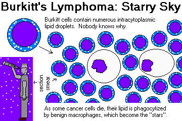
The "starry sky" appearance of Burkitt's is a favorite exam question. Just to confuse you, tingible body
macrophages appear as similar "stars" against the not-so-blue-as-Burkitt's
"sky" of a normal lymph node.
Despite "Big Robbins", the stars of Burkitt's are more conspicuous than other
tingible-body macrophages because
they are heavily laden with lipid.
{23620} Burkitt's lymphoma, lipid drops
{46336} Burkitt's lymphoma, lipid drops
{23611} Burkitt's lymphoma, good starry sky
{46332} Burkitt's lymphoma, good starry sky
{23641} * Burkitt's, methyl green pyronine (the red "pyroninophilia" merely tells us that the cytoplasm
is rich in ribosomes)
{46326} African Burkitt's, tonsils
AFRICAN BURKITT'S, almost always EBV-positive, is generally curable with chemotherapy, if you can get it to the victims.
By contrast, AMERICAN BURKITT'S, a sporadic disease of young people
and the immunocompromised, may be
EBV-positive or EBV-negative.
It can produce masses most anywhere, and has a worse prognosis.
MYCOSIS FUNGOIDES / SÉZARY SYNDROME (Lancet 371: 945, 2008.
Lymphomas of the epidermis and upper dermis, composed of large T4-cells with very elaborately
infolded ("cerebriform") nuclear membranes. The distinctive "Pautrier microabscesses" (misnamed)
are clusters of these T-cells within the epidermis.
In "mycosis fungoides" (Latin for "Toadstools! Toadstools!"), patients suffer from red, peeling skin for
some years, then enter a plaque and eventually a tumor phase, in which the patient looks horrible and
has lymphoma throughout the body.
{40003} mycosis fungoides
{40004} mycosis fungoides
{12747} mycosis fungoides, plaque phase
{12751} mycosis fungoides
{12754} mycosis fungoides
{13117} mycosis fungoides
{13781} mycosis fungoides
{13784} mycosis fungoides
{24740} mycosis fungoides, histopathology; note Pautrier microabscesses
{12759} mycosis fungoides, Pautrier microabscesses
{13793} mycosis fungoides, Pautrier microabscess
{13796} mycosis fungoides cells in a lymph node (look
how wiggly the nuclear membranes are)
{09042} mycosis fungoides cell, electron micrograph
In "Sézary syndrome", the red skin does not transform into tumors. Instead, the cells circulate in the
blood as a leukemia. The disease is slowly progressive, and survival for several years is usual.
{12757} Sézary patient
{16544} Sézary cell
{23722} Sézary cell
{15409} Sézary cell
* These are still probably incurable. Vorinostat, the novel chemotherapeutic
agent that inhibits acetylation of nucleosomes (?!), was approved in 2006
and seems to work nicely for otherwise-intractable T-cell cutaneous
lymphoma. See Blood 109: 31, 2007.
ADULT T-CELL LEUKEMIA-LYMPHOMA
A rare, very aggressive malignancy of T-helper cells.
It is strongly linked to the HTLV-I retrovirus, which is transmitted like AIDS, binds to the same
receptor (CD4), is neurotrophic, and lies dormant for a long time. (All about
HTLV-1: Lancet 353: 1951, 1999).
We now check all donor blood for this virus.
* The malignancy is preceded by polyclonal T-cell hyperplasia, due to induction of T-cell IL-2 receptors
by the virus.
* Have a pathologist show you the distinctive "flower cell" lymphocytes in
the blood in the leukemia.
* Hypercalcemia is common in this disease; the molecular biology
is curious, and involves the leukemic cells turning into osteoclasts (Blood 99: 634, 2002).
The disease (like the virus) is more common in Japan and the Caribbean. HTLV-I in Japan: Lancet
343: 213, 1994.
* Don't worry about the cancers of monocyte-macrophage origin.
MALIGNANT HISTIOCYTOSIS ("histiocytic medullary reticulosis"), a very aggressive, fortunately rare cancer
of blood-cell-eating true macrophages, is worth mentioning here.
Not a cancer, but also deadly....
HEMOPHAGOCYTIC LYMPHOHISTIOCYTOSIS,
a sometimes-genetic (often perforin), sometimes-acquired (triggered by infection or rheumatologic disease: Pediatrics 118: e216, 2006;
Blood 106: 3090, 2005;
South. Med. J. 100: 208, 2007) illness. The macrophages go crazy and destroy the body
(Am. J. Clin. Path. 137: 786, 2012).
{23668} malignant histiocytosis with erythrophagocytosis
TREATING THE NON-HODGKIN'S LYMPHOMAS:
This is HUGE news. We've already mentioned some of the novel agents
that are working. Rituximab "Rituxan", an antibody against CD20, is revolutionizing
treatment of B-cell lymphomas (Am. J. Clin. Path. 119: 472, 2003;
Blood 101: 949, 2003; NEJM 366: 2008, 2012; lots more).
Also watch for 90-Yttrium-ibrituxomab tiuxetan (Blood 99: 4336, 2002;
Cancer 94: 1349, 2002). Perhaps most exciting so far: I131-tositumomab, radioactive anti-CD20,
with a high five-year symptom-free survivals in follicular lymphomas.
Much success reported for indolent diffuse non-Hodgkin's lymphoma with
chemotherapy plus radioimmunotherapy (90-Yttrium-ibritumomab tuixetan: Cancer 112:
856, 2008).
Update on about a dozen monoclonals for B-cell lymphoma: NEJM 359: 613, 2008.
Claim of cures with non-myelogenic ablative transplantation (with or without 90yttrium ibritunonab tiuxetin):
Blood 119: 6373, 2012.
Future pathologists: In patients treated with rituximab who perish,
you'll see profound depletion of normal B-cells throughout the body
for as long as a year after treatment (Am. J. Clin. Path. 130: 604, 2008).
HODGKIN'S DISEASE ("Hodgkin's lymphoma"; J. Clin. Path. 55:
162, 2002)
A common (7500 cases/year in the U.S.), usually-curable cancer that typically affects young adults.
(There is a second peak in older adults; their disease tends to be more aggressive.)
Risk factors are ill-defined, and "epidemics" could perhaps be statistical accidents. Family members
are at several times increased risk, and a monozygous twin is at 100 times the base risk (NEJM 332:
413, 1995).
A previous history of Epstein-Barr infectious mononucleosis
supposedly triples one's risk for Hodgkin's
disease. This has held up and having Epstein-Barr virus on board
places one at an increased risk, but exactly what the relationship is remains
obscure (possible mechanisms: Blood 106: 4345, 2005). Of course,
many Hodgkin's patients are EBV-negative (Blood 106: 2444, 2005).
* Hodgkin's disease is rare in the Orient. For some reason, pediatric Hodgkin's is common in the
poor nations.
* Hodgkin's disease is a recognized complication of AIDS, though less typical
than non-Hodgkin's lymphomas. Not surprisingly,
AIDS patients with Hodgkin's disease tend to lack lymphocytes (Cancer 67: 1865, 1991).
The malignant cell is the REED-STERNBERG CELL, but until the late stages of the disease, the tumor masses
are composed primarily of inflammatory cells responding to the cancer.
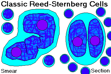
You must recognize the CLASSIC REED-STERNBERG CELL:
- 15-45 microns across
- multilobed nucleus (often appears "binucleate"), with lobes appearing as mirror images of one
another
- large, red owl-eye nucleoli, surrounded by clear nuclear sap
- pink-to-lavender cytoplasm
- CD15 positive (except nodular lymphocyte predominant subtype)
| |
|
{23560} Reed-Sternberg cell
{20057} Reed-Sternberg cell
{36398} Reed-Sternberg cell, not H&E; cytology
{36401} Reed-Sternberg cell, not H&E; cytology
{40423} Reed-Sternberg cell, mitosis
Everybody accepts the 1965 RYE CLASSIFICATION of Hodgkin's disease
with the W.H.O. modification that separated-out nodular lymphocyte predominant.
NODULAR LYMPHOCYTE PREDOMINANT:
In this illness, there are sheets of
lymphocytes and some RS-like cells.
However, the RS cells don't even immunostain like in other forms of Hodgkin's (they
are CD15-,
CD45+, B-cell markers are positive), and it's
probably "not really Hodgkin's, maybe a dysplasia":
old work Blood
87: 2428, 1996;
Am. J. Path. 146: 812, 1995; different mutations Blood 101: 706, 2003;
long-term follow-up supports the good prognosis but a few patients transform to a
diffuse large B-cell lymphoma (Cancer 116: 631, 2010).
The main reason to "type" Hodgkin's is to rule this in or out, since it's noted
for late recurrence (NEJM 318: 214, 1998).
CLASSICAL LYMPHOCYTE PREDOMINANT: A background of normal, monotonous, small lymphocytes.
{46338} Lymphocyte predominant Hodgkin's
{46339} Lymphocyte predominant Hodgkin's
Reed-Sternberg cells of any kind may be rare!
See NEJM 319: 246, 1988.
This variant generally announces itself in a single group of nodes, and almost all patients get cured by
today's therapies.
Don't diagnose "chronic lymphocytic leukemia" or "small lymphocytic lymphoma" in a young person
until you've sectioned through the block in your search for the diagnostic cell.
MIXED CELLULARITY: There are many Reed-Sternberg cells and variants, in a background of lymphocytes,
plasma cells, eosinophils, and histiocytes. This variant can present at any stage.
{23539} mixed cellularity Hodgkin's disease
{46342} mixed cellularity Hodgkin's disease
{46343} mixed cellularity Hodgkin's disease
{23524} lymphocyte depleted Hodgkin's disease. Just plain anaplastic.
{23542} nodular sclerosing Hodgkin's disease
{23545} nodular sclerosing Hodgkin's disease
{23548} lacunar Reed-Sternberg variants
{23551} lacunar Reed-Sternberg variant
NOTE: There are subtypes of each common type....
Sex ratios: Nodular sclerosis is a bit more common in women. All the other forms are more common
in men.
Having described this elegant classification scheme, I am almost sorry to have to add that the prognosis
for any particular case of Hodgkin's disease is determined by stage, rather than by type. Almost all
patients with stage I or IIA disease are now cured. This drops to around 50% for patients presenting
at stage IV.
Lymphocyte predominant presents at low stage, mixed cellularity at low or high stage, lymphocyte
depletion presents at high stage, and nodular sclerosis is often a mediastinal mass. These differences
account for "different prognosis for different Hodgkin's types".
* Update on treating Hodgkin's, including recognizing people with good prognosis for whom
the most intensive therapies can be avoided (Blood 120: 822, 2012).
REED-STERNBERG VARIANTS are also malignant.
MONONUCLEAR REED-STERNBERG-LIKE CELLS
("Hodgkin cells")
have single-lobed nuclei and one nucleolus. They may be seen
in any variant of Hodgkin's disease.
* LP CELLS (* "L&H cells") have scanty cytoplasm, big knobby nuclei, and small nucleoli. They are seen in lymphocyte
predominance Hodgkin's disease.
LACUNAR REED-STERNBERG CELLS have abundant, pale cytoplasm (* an artifact of formalin fixation). They
are seen in nodular sclerosis Hodgkin's disease.
POLYLOBULATED REED-STERNBERG CELLS ("popcorn cells") look like good Reed-Sternberg cells, except that the
nucleoli aren't so impressive. They are typical of mixed cellularity Hodgkin's disease.
* PLEOMORPHIC REED-STERNBERG CELLS are
anaplastic versions of the familiar form. They make up the bulk
of the tumor in lymphocyte depletion Hodgkin's disease.
REED-STERNBERG CELL RULES:
While a classic Reed-Sternberg-like cell may appear in other diseases (even "infectious mono"), its
presence in the proper background (see below) gives the diagnosis of Hodgkin's disease.
You must see a CLASSIC Reed-Sternberg cell before making the diagnosis.
Hodgkin's begins as an enlarged node or group of nodes. While we do not test you on staging,
everybody knows these basics (used for a majority of non-Hodgkin's lymphomas too):
Stage I... one node group or organ
Stage II... one side of the diaphragm
Stage III... both sides of the diaphragm
Stage IV...marrow, or two extra-lymphatic organs
"A" means no systemic symptoms
"B" means fever, weight loss (>10%), or night-sweats.
The classic Hodgkin's fever is the "Pel-Ebstein", or intermittent spiking fever.
{20056} Hodgkin's disease in a cervical node (we
would of course diagnose this only with microscopy)
{46348} Hodgkin's disease in the spleen
{46349} Hodgkin's disease in the spleen
Hodgkin's disease spreads predictably along contiguous groups of lymph nodes.
As it spreads, there may be transformation:
Lymphocyte predominant turns into mixed cellularity or lymphocyte depletion.
Mixed cellularity turns into lymphocyte depletion.
Nodular sclerosis generally keeps its type.
When I was in college, I had a classmate who I was told "might end up being
the first human cured of Hodgkin's disease." He died, but the fact that Hodgkin's is now cured so
often is one of the 20th century's great triumphs. Even chemoresistant Hodgkin's
now seems to be getting cured sometimes using a new protocol (with autologous stem cells, Cancer 113:
1344, 2008).
Minor mysteries of medicine:
(1) Hodgkin's patients often notice pain at sites of disease after they drink alcohol.
(2) Hodgkin's patients often have cutaneous anergy, even early in their disease.
INTRODUCING THE LEUKEMIAS
 Leukemia / myelodysplasia
"Pathology Outlines"
Nat Pernick MD
Leukemia ("white blood"), discovered and named of course by Virchow, is a generic term for replacement of the bone
marrow by cancerous blood cells. These usually (but not always; many acute leukemias are initially
"aleukemic") are spilling over into the bloodstream; in any case, expect a "packed marrow" except in
early CLL.
The story of progress in the treatment of the leukemias, all considered incurable when I was in college,
is described in Cancer 113(7S): 1933, 2008.
{23848} packed marrow; * this was late-stage CLL
{36032} packed marrow; * this was AML
{12347} packed marrow; * this was AML
ACUTE LEUKEMIAS ("poorly differentiated leukemias") are overgrowths of cells
that fail to mature (BLAST
CELLS). These diseases are very aggressive, and cause death in weeks or months.
{16243} blast with Auer rods
{29475} lymphoid blasts, pap stain. Big pale nuclei.
HOW TO SPOT A BLAST: (review from "Histology")
- scanty cytoplasm
- big nucleus with mostly euchromatin
- nucleoli
Future pathologists: You cannot tell whether a generic, undifferentiated "blast cell" is lymphoid or
myeloid without doing special stains and/or chromosomal studies
noted above. You'll learn below that Auer rods are sure markers
for myeloid differentiation. Of course, today you must use immunohistochemistry
(easy approach Arch. Path. Lab. Med. 132: 462, 2008).
* Auer rods in preleukemia: Am. J. Clin. Path. 124: 191, 2005.
These cells are not especially fast-growing, but they fail to mature. Even if they do not "crowd out"
their healthy neighbors, they tend to inhibit normal production of other blood cells.
Acute leukemia presents abruptly as one of the cytopenias (anemia, neutropenia, and/or
thrombocytopenia). Bone pain is likely to result from expansion of the marrow and infiltration of the
periosteum.
Later, involvement of other organs is common. Brain involvement is especially troublesome. T-cell
leukemias often produce a mass in the anterior mediastinum (why?).
{23842} acute lymphocytic leukemia, brain
{08734} acute leukemia, liver; as you would expect, the leukemia is blue
{08735} acute leukemia, liver
{08736} acute leukemia, liver
Death typically results from hemorrhage (cerebral, GI, other), and/or infection (neutropenia,
chemotherapy), and/or complications of bone marrow / stem cell transplantation.
{06269} fatal cerebral hemorrhage in leukemia
{01735} brain and dura, acute leukemia
The biology of the acute leukemias is very well-studied (still interesting Lancet 349: 118, 1997). The refractory ones
have pumps to remove chemotherapy drugs, etc., etc.
By contrast, CHRONIC LEUKEMIAS ("well-differentiated leukemias") feature cancer cells
that do mature
(more or less), and that have a natural history measured in years.
Like acute leukemia patients, these patients may present with a cytopenia problem. Or they may have
problems from a high white ("leukostasis" plugging small vessels), or may notice lymphadenopathy or
organomegaly, or the high white count may be an incidental finding on "routine lab".
RULE: Marrow cells (most leukemias, extramedullary hematopoiesis) in the spleen involve the red
pulp. Lymphomas in the spleen (at least the B-cell type) involve the white pulp.
RULE: Any leukemia can involve the lymph nodes and make them large, but the enlargement is seldom
so spectacular as in Hodgkin's or non-Hodgkin's lymphoma.
RULE: Any leukemia (or lymphoma) can involve the liver, but enlargement is usually not spectacular.
Hodgkin's and non-Hodgkin's lymphomas will first appear in the portal areas.
LYMPHOID LEUKEMIAS recall the various kinds of normal lymphocytes, and
MYELOID ("myelogenous",
or better, "granulocytic") leukemias recall a normal granulocyte.
You remember that MYELOCYTES, the precursors of granulocytes, are the most common cell in normal
bone marrow (MYELO-); hence their name.
Cell turnover in the leukemias (and the closely-related polycythemia vera and
primary myelofibrosis) is much increased, placing these patients at risk for gout. The risk increases further when
cancer cells are dying by the pound during therapy. The thoughtful oncologist gives prophylactic
medication.
ACUTE LYMPHOBLASTIC LEUKEMIA ("ALL"; diagnosis Am. J. Clin. Path. 111:
467, 1999)
This is the familiar "childhood leukemia", with peak age in four year old kids. Adult ALL is less
common.
* Authoritative mega-review for pathologists considering
a diagnosis of leukemia in a child: Am. J. Clin. Path. 109(4S1):9S, 1998.
{12410} ALL (all you can tell from the smear is "blasts")
ALL seems to strike at random. Radiation exposure is a known risk factor, Down's syndrome kids are
at 15x the normal risk, a patient's identical twin has a 20% risk.
* A curious claim that could be true is the idea that leukemia is an unusual response to one
or more as-yet-unidentified viruses met at the wrong age; one finding that seems to be robust is a
strong correlation between leukemia rates and the diversity of origins, i.e., new immigrants, in an area
(Br. Med. J. 313: 1297, 1996; Lancet 349: 344, 1997.)
Rachel Carson's unfortunate (i.e., false and inflammatory)
discussion of leukemia in "Silent Spring"
is now history. She was right about the danger of DDT to birds' eggs, and her
manipulation of the data on leukemia is a minor blot on the memory of a great science
writer. Nobody's perfect.
Today, there's no serious discussion of the old alleged link
between insecticide exposure and leukemia (* the one paper I could find from
the decade 1997-2007, Canc. Ep. 16: 1172, 2007, looks like
recall bias).
* Pathologists and oncologists used to subclassify
acute lymphoblastic leukemia by blast morphology
("FAB classification"; stands for "French-American-British"):
L1: 85%...
Cells <= 2x the diameter of a normal lymphocyte; smooth nuclei; more common in kids
L2: 14%...
Bigger cells, lots of clefts, often nucleoli; more common in adults
L3: 1%...
Even bigger cells, Liquid Burkitt's lymphoma; t(8;14) and everything
{23746} L1
{23833} L1, special stain (cytoplasm is brown)
{23860} L2
{13982} L2, bone marrow
{23758} L3; note the lipid
{13985} L3
L3 is a distinct entity, but L1 and L2
aren't especially useful. Here's a more-modern immunophenotypic classification:
B-cell... 80%... * CD19+...* several subclasses exist
"pre-B"/"null", with surface Ig and TdT, carries a good prognosis;
Burkitt's / "mature B" / L3 is ominous
T-cell... 15%... * "intra-thymic markers, like T-lymphoblastic lymphoma"; chances of a cure are much less than with
B-cell disease
Nothing... uncommon today...* markers are negative; there is only HLA-DR.
Apart from the fact that all L3's (Burkitt's) are B-cell tumors, there is little correlation between the two
systems.
In 2002, the World Health Organization proposed the following system, which seems to be generally accepted now:
More for cytogeneticists and prognosticators:
Hyperdiploidy (especially "high hyperdiploidy", >50 chromosomes, all good standard ones), present in a large minority of B-cell ALL's, confers a good
prognosis.
* Fans of crackpot science please take note: If the
"independent thinker / persecuted genius" claim that cancer is not caused
by mutations but by nondisjunction, this curious fact about leukemia would not be true.
Adults with ALL are often "Philadelphia-positive", and kids can be, too (* the latter
confers a bad prognosis: NEJM 342: 998, 2000). Several other
bad-prognosis translocations, including the Burkitt t(8:14), and the AML
variant of the Philadelphia chromosome, are known.
t(12;21) and del(9p) give a relatively favorable prognosis.
Almost all children with ALL get a complete remission on current therapy. Around
90% get apparent cures on today's regimens (NEJM 366: 1445, 2012).
The prognosis for adults is still guarded. Cure rates for teens
are now about as good as for young children (J. Clin. Onc. 29: 386, 2011).
Pccasionally the disease appears in older adults with less chance of cure.
* Precursor T-cell ALL and MLL gene-rearranged leukemias
are exceptions that are more refractory to cure.
{23857} ALL in the liver
{32027} ALL in the liver
{34513} ALL in the brain
{49316} ALL in the kidney
{49343} ALL in the testis
* In 1987, Coby Howard,who had ALL and a relapse, and belonged to a family
that did not work and received Medicaid as part of welfare,
didn't get his $100,000
bone marrow transplant that had maybe 25% chance of working (J. Clin. Onc. 5: 1348, 1987 is from that era)
because of Oregon's
health-care rationing, which was intended to spend
the Medicaid money where it was most likely to do the
most overall public good.
Pressure groups (liberal, conservative), of course, had a field-day with Coby's death.
During the activity at the legislature that followed, it was pointed out that
many working people who did not have health insurance were dying because
they lacked access both to transplants and to far more basic services.
Taxpayers understandably resent being forced to pay for health benefits for others
that they
cannot get for their own children.
Remember
poor Coby's name, and the principle.
ACUTE MYELOID LEUKEMIA ("AML", "acute myelogenous leukemia", "poorly differentiated granulocytic leukemia",
"acute non-lymphocytic leukemia", "ANLL", etc.)
This is the common acute leukemia of adults (the incidence increases with age, but young adults are often
affected, and occasionally children are affected).
Although most cases are idiopathic, many things are known to increase risk. These include:
- Down's syndrome (trisomy 21 -- especially AML-M7)
- those fragile chromosome syndromes
Bloom's
ataxia telangiectasia
Fanconi's anemia (a complex genetic disorder; * gene
FancC Blood 94: 1, 1999; leukemias Cancer 97: 425, 2003.)
- benzene exposure (* supposedly the metabolites inhibit a topoisomerase: Blood 98: 830, 2001)
- ethylene oxide exposure
- ionizing radiation exposure ("we estimate one case per 10000 childhood CT scans" -- Lancet 380:
499, 2012).
- previous cancer chemotherapy (NEJM 322: 52, 1990 is still good; remember the alkylating agents and anthracyclines; the
risk from adjuvant breast cancer therapy is minimal if any Cancer 101: 1529, 2004)
- any "myelodysplastic syndrome" ("preleukemia")
- any "chronic myeloproliferative syndrome" ("blast crisis" -- * many of these patients
enter "leukemia transformation" by acquiring a mutated SRSF2; see Blood 119: 4480, 2012)
- primary myelofibrosis
- polycythemia vera rubra
- "essential" hemorrhagic thrombocythemia
- chronic myelogenous leukemia
- "idiopathic aplastic anemia" (usually T-cell autoimmune)
The cancer arises out of a background of mutated marrow cells that typically include granulocytic
and other (erythroid and/or monocyte precursors). All are likely to show some abnormalities (* good
tipoff: megakaryocytes with non-lobulated nuclei).
The FAB classification (Ann. Int. Med. 103: 614 & 620, 1985) is worth knowing at the recognition
level (though it is of little interest to practicing oncologists, who are today much more concerned about
the molecular lesions):

M0: "minimally differentiated AML"; undifferentiated myeloblasts without myeloperoxidase (* new criteria Am. J. Clin. Path. 115: 876, 2001)
M1: "acute myeloblastic leukemia without maturation"; undifferentiated myeloblasts with myeloperoxidase; just maybe a few Auer rods
M2: "acute myeloblastic leukemia with granulocytic maturation"; the most common; some promyelocytic differentiation, maybe a few Auer rods; * t(8;21) is distinctive, with fusion AML1/ETO
M3: "acute promyelocytic leukemia"; very granular promyelocytes, often many Auer rods, DIC
(* from annexin II on the surfaces that activates plasmin:
NEJM 340: 944, 1999; not the full story Thromb. Res. 113: 109, 2005); * t(15;17)
and * t(11;17) are distinctive, and (KNOW:)
involve the vitamin A receptor (RARα; discovery Blood 77: 1418 & 1657,
1991).
All-trans retinoic acid
(* "tretinoin") matures these cells: NEJM 324: 1385, 1991;
NEJM 27: 385, 1992;
Blood 85: 2643, 1995; Blood 94: 39, 1999
(* discovered by the Mainland Chinese, who attributed their success to lack of
regulation of human research.... Do any of our "ethicists" want to "refuse to use this data which was
acquired immorally"?)
* Retinoic acid plus G-CSF for the difficult t(11;17) subtype:
Blood 94: 39, 1999.
Diagnostic stain Blood 86: 862, 1995.
Arsenic trioxide (!) was introduced into the treatment of M3 (first positive report
NEJM 340: 1043, 1999. Taken in combination with the retinoic acid derivatives,
the new protocols produce cures (Nat. Med. 14: 1333, 2008; NEJM 360:
928, 2009.)
M4: myeloid and monocytic differentiation (* a subtype M4eo has eosinophilis all over the marrow)
 Acute Myelomonocytic Leukemia
Text and photomicrographs. Nice.
Human Pathology Digital Image Gallery
M5: monocytic differentiation only; * t(9;11)(p22;q23) is typical of M5a; 9p22 is beta-interferon receptor, while 11q23
is the MLL
(myeloid-lymphoid leukemia gene; common
in infant and adult leukemias, though not childhood leukemias
(Proc. Nat. Acad. Sci. 88: 10735,
1991; Blood 94: 283, 1999) as well as adult AML
* MLL also forms a fusion product with MEN-1 (which
is adjacent) and a src-like gene on chromosome 19 (Proc. Nat. Acad.
Sci. 94: 2563, 1999) in t(11;19)
leukemias; this fusion product inhibits p53's product (Blood 93:
3216, 1999).
* M5a is "acute monoblastic leukemia" with very primitive-looking blasts
bearing monocyte markers; M5b is "acute monocytic leukemia" with a dented
nucleus and granules.
M6: "acute erythroid leukemia"; features of red cell precursors predominate; "Di Guglielmo's erythroleukemia" (* future
pathologists: look for d-PAS-positive chunks in the cytoplasm)
M7: "acute megakaryoblastic leukemia"; platelet markers; acute marrow fibrosis (* PDGF effect; reticulin);
acute megakaryoblastic leukemia is a childhood leukemia (Am. J. Clin. Path. 122-S: S33, 2004).
Rarities not in the schema include acute basophilic leukemia, acute eosinophilia, and several others.
The World Health Organization's 2002 attempt to reclassify the acute myelogenous leukemias
in terms of their mutations and clinical antecedents (Blood 100: 2292, 2002)
met with mixed reviews; you will
find it in research papers. A new version came out in 2008.
- AML with a characteristic genetic abnormality (several are listed; generally better prognosis)
- AML with multilineage dysplasia (i.e., arising from myelodysplastic syndrome, polycythemia vera, myeloid metaplastia, etc., etc.; WHO claims poor prognosis but this is disputed Blood 1111: 1855, 2008)
- AML resulting from previous radiation or chemotherapy (generally poorer prognosis)
- AML not otherwise categorized -- unfortunately, a very long list based on morphology
- "Acute leukemia of ambiguous lineage" -- both T-cell and B-cell markers, or neither T-cell nor B-cell markers
Researchers expressing ongoing dissatisfaction with the WHO 2002 system (for example, the
fact that 40-50% of adults with AML have no cytogeneic abnormality at diagnosis)
are looking forward to a future classification based on individual mutations, instead.
* Watch FLT3 (a tyrosine kinase), nucleophosmin (NPM1 -- the most-often
mutated gene in AML
and now stainable in some normal-karyotype AML's: Am. J. Clin. Path. 133: 34, 2010; now used as a guide
to treatment and to screen for residual disease Arch. Path. Lab. Med. 135:
994, 2011), "myeloid/lymphoid mixed-lineage
leukemia gene" (MLL) and mutations and overexpressions in numerous others.
Studies are now being designed focusing on different treatments depending on
these mutations.
ALso often mutated rather than translocated in AML is DNMT3A, which makes the
prognosis more ominous (NEJM 363: 2424, 2010.
Update on the genetics of acute myeloid leukemia: NEJM 366: 1079, 2012
Be all this as it may, we begin making the distinction
among the many variants of AML from one another, and from ALL, by morphology and by the stains
listed earlier in the unit. AUER RODS are red-staining, rod-shaped crystalloids made of primary granules
(you can see them especially well if you stain for peroxidase).
If present, they prove that a blast is "myeloid" and not "lymphoid".
{12338} AML. Note promyelocyte in center.
{16245} blast, and not much else, M1
{13988} blasts, and not much else, M1
{23830} blast, * chloroacetate-esterase (+), M1
{13940} blast, * chloroacetate-esterase (+), M1
{23839} blast, M1; note nucleolus.
{23770} blasts with a few granules, M2
{13991} blasts with a few granules, M2
{16249} blast with Auer rod, M2
{12344} blast with Auer rod, M2 or maybe M3
{14010} blast with great Auer rods
{23776} blasts with lots of granules, M3
{13994} blasts with lots of granules, M3
{16254} blasts with lots of granules, M3
{10109} blasts with lots of granules, M3
{13997} semi-monocyte like blasts, M4; note indented nucleus and gray cytoplasm
{10133} semi-monocyte like blasts, M4
{13937} non-specific esterase in M4
{23800} monocyte-like blasts in M5
{40449} monocyte-like blasts in M5
{40451} monocyte-like blasts in M5
{23803} non-specific esterase in M5
{14001} monocyte-like blasts in M5
{13928} non-specific esterase in M5
{23949} erythroleukemia, M6. If you don't recognize the malignant cells as red-cell precursors, please
check out a histology book.
{14007} erythroleukemia, M6
{16267} erythroleukemia, M6
{23836} erythroleukemia, M6
{23806} erythroleukemia, M6
{16273} erythroleukemia, M6, PAS-positive chunks
{16274} megakaryocytic blasts, M7. See the platelets budding?
{23812} megakaryocytic blasts, M7
{23821} megakaryocytic blasts, M7
{23818} megakaryocytic blasts, PAS-positive, M7
{46340} gingival involvement; this is common in M4 & M5

Untreated AML is fatal in a few weeks.
Today, many AML patients have prolonged survival and probably
cures.
The prognosis depends primarily on the cytogenetics. The major papers
came from the early 2000's (Blood 96: 4075, 2000; Blood 100: 4325, 2002).
"Favorable prognosis"
features t(8:21), t(15:17), del(9q) and/or inv(16)/t(16;16).
"Adverse prognosis" features -5 / del(5q), -7 / del(7q), -17, -18, inv(3)/t(3;3),
t(6;9), t(9;22)
and/or complicated cytogenetics (i.e., three or more unrelated abnormalities).
The others, including those with
no chromosomal abnormalities, are "intermediate prognosis."
Acute promyelocytic leukemia has the best response to treatment,
with 98% adjusted six-year survival (Blood 110: 59, 2007).
A solid growth of myeloblasts is a CHLOROMA or GRANULOCYTIC SARCOMA
or NONLEUKEMIC MYELOID SARCOMA. This most
malignant of solid tumors turns green (Greek "chloros",
as "chlorine" or "chlorophyll") on exposure to air (why?)
It's not common, but is treated as, and responds similarly to, AML, with a
somewhat better chance of cure (Cancer 113: 1370, 2008).
TRANSIENT MYELOPROLIFERATIVE DISORDER ("transient leukemia")
is seen in the first weeks of life in around 10% of children with Down's trisomy
21. The white count rises very high with many blasts (tending to be knobby,
like megakaryoblasts, or else to resemble minimally differentiated AML), and platelets are likely to
be high. There are usually not many blasts in the marrow,
but there's a tendency to involve the liver. It usually self-cures, but these children are at risk for later acute myeloid / megakaryocytic
leukemia (signature mutation in both conditions in GATA1: Lancet 361:
1617, 2003; Blood 101:
4301, 2003; Blood 102: 2960, 2003; J. Ped. 687, 2006; Blood 111:
2991, 2008); the occasional
child without Down's who gets transient myeloproliferative disorder in the
nursery is at similar risk.
THE MYELODYSPLASTIC SYNDROMES ("THE PRELEUKEMIAS", Carl Sagan's disease):
Mayo Clin. Proc. 70: 673,
1995; Am. J. Clin. Path. 119(S1): S-58, 2003.
This is a family of disorders in which there are problems in producing red cells, granulocytes, and
platelets. What has happened is that the normal precursor cell that gives rise to all three has been
replaced by a mutant clone.
Various karyotypic abnormalities have been described in the majority of
these people (Virch. Arch. A 421: 47, 1992).
* A 5q- predicts a good response to the
thalidomide analogue lenalodomide (NEJM 352: 549, 2005).
* Isochromosome 17q, still mysterious, is a
common lone abnormality (Blood 94: 225, 1999); my late mother,
who received radiation as a child for osteomyelitis, had Ph'-negative CML
with just this
signature;
this is also a common known hit in CML's progression to
blast crisis.
Unlike most of the other neoplasms of white cells,
several genes are known in which point mutations
confer an unfavorable prognosis (NEJM 364: 2496, 2011).
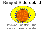
* Subclassification is useful. The old FAB classification:
1. Refractory anemia (poor hemoglobinization, too few red cells)
2. Refractory anemia with ringed sideroblasts (>15% of nucleated red cells)
3. Refractory anemia with excess blasts (5-20% myeloblasts)
4. Refractory anemia with excess blasts in transformation (20-30% myeloblasts)
5. Chronic myelocytic leukemia
Ask your hematologist what this all means. The World Health Organization
put out a new, more-elaborate clasification with criteria in 2002 (Blood 100: 2292, 2002)
and tweaked it in 2008 with instructions on how to distinguish cell types; leave it to us.
* Somatic mutation in the ringed-sideroblast form SF3B1: NEJM 365: 1384, 2011).
Most patients are older adults, who sometimes are symptomatic. In the more aggressive forms, death
follows in a few years. Often these patients are asymptomatic, and the problem is detected on routine
screening.
As you would expect, this disease pattern has a propensity to transform into
acute myeloid
leukemia, and does so in a large minority of cases.

 Myelodysplastic syndrome
Odd megakaryocyte / giant platelet
AFIP
CHRONIC MYELOID LEUKEMIA ("chronic myelogenous leukemia", "well-differentiated
granulocytic leukemia"): all about it Lancet 370: 1127, 2007
This is cancer of the myeloid stem cells in which there is overgrowth of normally-maturing myeloid cells
Radiation and exposure to chemicals (notably benzene) are known risk factors. Most of the time, the
disease seems to strike at random.
Patients typically have high counts of neutrophils and their precursors (and almost always basophils).
These are normal (functionally and morphologically) for all intents and purposes, except that for some
reason they lack cytoplasmic alkaline phosphatase.
Preposterously high white counts (>100,000 or so) are likely to result in white cells plugging important
small vessels ("leukostatic ischemia" of the brain, etc.)
While the spleen is likely to be enlarged in all the common leukemias, chronic myeloid leukemia
typically produces huge spleens (down almost to the pubic hair). There will usually be some little
infarcts.
Occasional CML cases have predominance of basophils (itchy) or eosinophils. Serum vitamin B12 is
likely to be elevated due to elevation of its binding protein; this can happen in other myeloproliferative
disorders.
{10769} CML, splenomegaly
{10763} CML, peripheral blood
{12359} CML
{23863} CML (note the basophil)
{23866} CML,
leukocyte
alkaline phosphatase stain (black; note the cells are not stained black)
{12371} Philadelphia chromosome
 Chronic granulocytic leukemia
Philadelphia chromosome
WebPath Photo
The disease eventuates, after a few years, in BLAST CRISIS, with or without (50%/50%) a previous
accelerated phase. Blasts appear in the circulation in large numbers (30% or more), and death follows quickly as they
overwhelm the marrow and body. This is not very treatable.
* Determining the clinical phase of CML by labs: Cancer 106: 1306, 2006.
"Accelerated phase" has begun when there are 10-29% blasts or >30% blasts-and-promyelocytes
in blood or marrow,
persistently large spleen, lots of basophils, big spleen or thrombocytosis unresponsive to therapy,
or thromcytopenia unresponsive to therapy.
{23869} CML, blast crisis
{12365} CML, blast crisis
Traditional chemotherapy with busulfan or hydroxyurea
controls symptoms during the chronic phase but neither
speeds nor delays blast crisis.
Newer therapies (alpha-interferon with or without cytarabine)
suppress the leukemic clone and do prolong survival.
Historically, the only hope for a cure was allogenic bone marrow transplantation.
Watch the outcome of people treated long-term with the new biotech products.
In around 30% of these cases, the blasts express lymphoid differentiation (TdT, etc.) T-cell blast crisis:
Am. J. Cln. Path. 107: 168, 1997.
WARNING: The following "myeloproliferative diseases" are all "tumors of the multipotent myeloid
stem cell", and can transform into one another (usually from a mild one to a bad one):
polycythemia vera rubra
hemorrhagic ("essential") thrombocythemia
primary myelofibrosis
idiopathic "aplastic anemia"
chronic myelogenous leukemia
"APLASTIC ANEMIA" (updates Ann. Int. Med. 136: 534, 2002;
Lancet 365: 1647, 2005; Blood 110: 1603, 2007)
This uncommon, dread illness features the disappearance of the precursors of
granulocytes, erythrocytes, and platelets. (It is poorly-named.)
In the 1990's, it became clear that most cases of aplastic anemia are caused
by T-cell-mediated attacks on the hematopoietic marrow. This explains why...
a direct marrow / stem cell transplant from an identical twin never takes in this disease;
- bone marrow / stem cell transplant after anti-thymocyte treatment is often curative
- anti-thymocyte globulin plus cyclosporine restores marrow function to most of these patients, most often permaently (JAMA 289: 1130, 2003)
- children with severe combined immunodeficiency who get graft-vs.-host disease from a blood transfusion develop aplastic anemia
A key to the autoimmunity in aplastic anemia may have been
discovered. Many of these people have a clone of T-cells
with mutated perforin (Blood 109: 5234, 2007), the same
locus that's mutated in familial hemophagocytic lymphohistiocytosis.
Other cases of acquired aplastic anemia
seem to be the result of running out of telomere length
during aging. Some families and some individuals
have less telomerase than others (to oversimplify, but
the impact seems real): NEJM 352: 1413, 2005.
Once uniformly fatal, the disease is now often controllable using
marrow / stem cell transplantation and/or immunosuppression (Blood 108: 2509, 2006).
 Aplastic anemia
WebPath Photo
CHRONIC LYMPHOCYTIC LEUKEMIA ("CLL", "well-differentiated lymphocytic leukemia"; "the
liquid phase of well-differentiated lymphocytic lymphoma", etc.) Lancet 371: 1017, 2008.
This indolent cancer is a clone of B-cells that multiply slowly and do nothing useful.
The diagnosis is made by finding a count of 5000 or more lymphocytes of appropriate
phenotype circulating in the blood.
If the cells have nucleoli, it's more likely to be called
B-cell prolymphocytic leukemia and to behave more aggressively.
Often you can find growth centers (where the cells are slightly larger
and perhaps show nucleoli) in solid-phase well-differentiated lymphocytic
lymphoma; these have the same molecular markers as classic CLL.
"T-cell CLL", with a peripheral smear looking like CLL,
is now renamed "T-cell prolymphocytic leukemia", an uncommon
and aggressive disease. There are of course T-cell receptor
rearrangements, and most often inv 14(q11;q32).
The one known risk factor is ataxia-telangiectasia (homozygotes,
very likely heterozygotes), and not
surprisingly, this gene is often mutated in sporadic cases
(Lancet 353: 26, 1999.)
We know of no other specific risk factors, not even a history of radiation.
* CLL has a few common genetic markers but they also occur in other B-cell neoplasms
(Semin. Hem. 36: 171, 1999.
- del 13q14 is common; nobody knows the deleted anti-oncogene.
- t 11;14, the bcl1 gene meets IgG, is also common
- Many cases of B-cell CLL have trisomy 12 (you can see this in a variety
of other B-cell problems.)
- A few others have turned up recently. Stay tuned (NEJM 365: 2497, 2011).
The disease is often an incidental finding, when a CBC shows preposterously a high lymphocyte count.
In many of these patients, vimentin is lacking in the
cytoskeleton of the neoplastic cells.
Hence these cells are fragile on smears. Crushed CLL cells are called "smudges",
and it now seems that the higher percentage of smudges, the better the prognosis
(Mayo Clin. Proc. 82: 449, 2007).
{08784} CLL
{12389} CLL
{12404} CLL going bad (some blasts)
{12386} CLL with smudges
Paraneoplastic syndromes are more troublesome in this disease than in most other leukemias.
Around 15% of patients get autoimmune hemolytic anemia.
The lymphocytes do somewhat suppress the heathy plasma cells, and the patients have troubles with
infections.
* A few percent develop a marker paraprotein, usually kappa IgM, or get mu heavy chain disease.
Patients with anemia or thrombocytopenia from CLL survive around 2 years. Asymptomatic people
with CLL as an incidental finding generally survive more than ten years.
A few percent of patients develop a diffuse large-cell lymphoma. This is the rapidly-fatal
RICHTER'S SYNDROME (NEJM 324: 1267, 1991); predisposing
factors remain mysterious (Cancer 67: 997, 1991).
* PROLYMPHOCYTIC LEUKEMIA is an uncommon, aggressive variant of CLL.
Chemotherapy for end-stage CLL: NEJM 330: 319, 1994. No miracles.
* It will not surprise you to learn that many people have "pre-CLL", or
"monoclonal B-cell lymphocytosis", detectable only by zealous search for the mutated
cells
by pathologists. Each year there's only about 1% chance of transformation (NEJM 359: 638, 2008;
don't look for it Blood 119: 4358, 2012).
HAIRY CELL LEUKEMIA (Mayo Clin. Proc. 87: 67, 2012)
This distinctive leukemia is named for the many hair-like projections on its surface.
It was of unknown histogenesis (* confusing surface markers) until its molecular
genetics and antigenic markers
established it as a B-cell neoplasm" (Blood 104: 250, 2004; Am. J. Clin. Path. 125:
251, 2006).
Patients have circulating "hairy lymphocytes"; usually
have big spleens and are sometimes anemic, neutropenic, and/or thrombocytopenic.
Treatment is often unnecessary or can be delayed until the disease
gets symptomatic.
Bone marrow
aspiration is likely to be unsuccessful (dry tap) "because the cellular hairs tangle with one another"
(* more likely, because TGF-beta1 from the tumor induces extra reticulin fibrosis:
J. Clin. Inv. 113: 676, 2004.
The hairs are quite distinctive, and the diagnosis is clinched by the finding of
TARTRATE-RESISTANT ACID PHOSPHATASE (TRAP) in these cells.
* Future pathologists: The other "hairy" B-cell neoplasm is "lymphoma with
villous lymphocytes", usually in the spleen. The distinctions are subtle, and
the immunotyping of tumors is often variable. Arch. Path. Lab. Med. 124: 1710, 2000.
The genetics of hairy cell leukemia are finally
being discovered.
A trademark (probably driver) BRAF mutation (to the familiar BRAF V600E
known from other tumors) has just been found in each of 48
hairy-cell patients (NEJM 364: 2305, 2011; assays Blood 119:
3151, 2012; Am. J. Clin. Path. 138:
153, 2012).
During the 20th century, the only treatment for this disease was splenectomy, which helped.
Today, most patients get a lasting remission
after taking a course of cladribine (2-chlorodeoxyadenosine, 2-CdA) or pentostatin
(deoxycoformycin, a purine analogue that's a naturally-occurring antibiotic).
Update Cancer 104: 2442, 2005; Blood 109: 3672, 2007.
These can be repeated is required if the disease recurs (which it often
doesn't), and there are additional treatments that give results if the
disease becomes resistant.
* There is a variant that features mutated p53 instead of BRAF V600#
and is much
more common in men and is harder to treat. There's also a Japanese
variant that responds very well to cladribine.
{23872} hairy cell leukemia
{10766} hairy cell leukemia, spleen (top; normal at bottom)
{16543} hairy cell leukemia, TRAP stain (red)
{23875} hairy cell leukemia, TRAP stain (red)
{13925} hairy cell leukemia, TRAP stain (red)
{16541} hairy cell leukemia, TRAP stain (red)
{23881} hairy cell leukemia, bone marrow biopsy (trust me)
{42117} big spleen in hairy cell leukemia, foot ruler
POLYCYTHEMIA VERA ("Osler's polycythemia", "P. V. rubra", etc.; Mayo Clin. Proc. 78: 174, 2003; Arch. Path. Lab. Med. 130: 1126, 2006)
By convention, POLYCYTHEMIA (a better synonym is ERYTHROCYTOSIS) describes an abnormally high
hemoglobin. Classification:
ABSOLUTE POLYCYTHEMIA (i.e., increased circulating red cell mass):
PRIMARY POLYCYTHEMIA (i.e., the main problem is with the red cells)
Polycythemia vera rubra
NOTE: Cancer of the normoblasts (i.e., AML-M6) isn't considered a polycythemia
SECONDARY POLYCYTHEMIA (i.e., the main problem is elsewhere)
Effective renal arterial hypoxia
Emphysema
Sleep apnea
Tetralogy of Fallot
Hemoglobins with too much oxygen affinity
For a first-person story of injectable bioengineered erythropoietin and
bicycle racing, including how athletes beat the tests by infusing
huge amounts of normal saline by vein moments before having
their hematocrits checked, see Sci. Am. 298(4): 82, 2008.
Genetic errors in erythropoietin production or sensitivity ("familial erythrocytosis", for example HIF2A: NEJM 358: 162, 2008)
Erythropoietin-producing tumors
Renal cell carcinoma
* Hepatocellular carcinoma
* Cerebellar hemangioblastoma (?!)
Anabolic steroid users
After kidney transplant (over-zealous proximal tubule produces erythropoietin)
Altitude (above about 10,000 feet for a long time? Expect problems.
Stroke risk: Stroke 26: 562, 1995. Pulmonary vein thrombosis:
Hum. Path. 21: 601, 1990.
"Primary familial polycythemia" / "congenital primary erythrocytosis" (truncated
erythropoietin receptor stuck in the "on" position;
(J. Clin. Invest. 102: 124, 1999)
* Erythropoietin-dependent polycythemias (altitude, post-transplant) can
be ameliorated using ACE inhibitors, which is puzzling: Lancet 359:
663, 2002.
RELATIVE POLYCYTHEMIA (i.e., dehydration)
Polycythemia vera is a proliferation of stem cells (again, the common precursors of red cells,
granulocytes, and megakaryocytes). This time, they are very erythropoietin-sensitive and mostly mature
into red cells.
The cells are the common ancestors of red cells, neutrophils, and megakaryocytes. Over the course of
years, these stem cells replace the normal stem cells of the marrow. Their progeny, however, are fully
functional. (Neutrophils even have normal alkaline phosphatase levels.)
In addition to a high red cell count, white cells and platelets are likely to be high.
On biopsy, expect to see a very hypercellular marrow, with all cell lineages increased. In the late stages,
there is often
marrow fibrosis ("burned out PVR", "postpolycythemic phase", * PDGF effect?) or replacement by blasts (transformation to acute
myelogenous leukemia -- still no good treatment Cancer 104: 1032, 2005).
The trademark mutation in JAK2 (NEJM 356: 444, 2007) that is usually present
(* JAK2V617F) is now famous. Mouse with the mutation -- Blood 120: 166, 2012.
This is a disease of older middle-age. Until the last stages, patients are troubled primarily by the
increased volume of hyperviscous blood.
This causes congestion of most organs ("the plethoric face", etc.)
More troubling, the stasis of gooey blood in the veins promotes clotting.
Or distended veins can rupture (GI bleeding, hemorrhagic stroke). Eventually these patients get platelet
problems, too, which does not help the bleeding tendency.
Minor mystery of medicine: Itching after taking a hot shower is very suggestive of PVR.
The mainstay of treatment for polycythemia vera is regular phlebotomy, to keep the red cell count down.
These patients' survival curves are nearly as good as normal folks.
For all three of the JAK2 diseases, the major killer
is the tendency of the disease to turn into acute myelogenous
leukemia.
The once-popular practice of giving these patients radioactive phosphorus resulted in a greatly increased
rate of transformation to acute leukemia, turning a not-so-bad, easy-to-control disease into a lethal,
untreatable one. Later, the same thing happened with trials of chemotherapy.
The traditional criteria for the diagnosis of PVR:
A1... Increased RBC mass (>=36 mL/kg; >=32 mL/kg; the math comes to a hematocrit of 52 for a man, 46 for a woman)
A2... Normal arterial PO2
A3... Splenomegaly
B1... Platelets greater than 400,000/L
B2... WBC >=12,000/L
B3... Leukocyte alkaline phosphatase over 100 in the absence of evidence of infection
B4... Elevated serum vitamin B12.
Make the diagnosis if:
(1) You have A1 + A2 + A3, or
(2) A1 + A2 + any two B's.
In 2007, the World Health Organization suggested
requiring two major criteria (hemoglobin >18.5 g/dL;for men, >16.5
g/dL for women, plus a functionally active JAK2 mutation.
Minor criteria are one of these -- characteristic bone marrow
morphology, too-low serum erythropoietin, and the ability of red cell colonies to
grow in tissue culture without erythropoietin.
* Thrombosis, the most troublesome aspect of this disease,
seems to be much less of a problem if patients are simply given low-dose aspirin
(NEJM 350: 114, 2004).
 Polycythemia
Text and photomicrographs. Nice.
Human Pathology Digital Image Gallery
PRIMARY MYELOFIBROSIS ("myelofibrosis with myeloid
metaplasia"; "agnogenic myeloid metaplasia";
"myelosclerosis"); JAMA 303: 2513, 2010; Mayo Clin. Proc. 87: 25, 2012
Primary myelofibrosis is a proliferation of neoplastic stem cells in the bone marrow (which merely becomes
hypercellular)
and the red pulp of the spleen (which enlarges greatly). For some reason, the marrow
tends to develop increased reticulin / collagenize, "burn out" and become fibrotic.
* The WHO criteria require (1) megakaryocyte proliferation and atypia and usually fibrosis
of the marrow;
(2) not being one of the other recognizable myeloprolifeative diseases;
(3) some clonal marker such as JAK2 or one of the others or at least not chronic inflammation or cancer;
(4) Two of the following: leukoerythroblastic smear; increased serum LDH;
anemia; palpable spelen. These are going to be discarded when the molecular signatures
are better-defined.
Historically, ionizing radiation and benzene exposure
are known risk factors.
We know it's tumor since there's a group of common translocations;
however, which ones are present doesn't impact prognosis at least much (Cancer 107:
2801, 2006). Most now seem to have mutated JAK2, and it seems likely this will
soon define the disease.
By definition, BCR-ABL (t(9;22), Philadelphia chromosome) is absent.
As in polycythemia vera, the cells that enter the blood are fully functional. This time, there is no
tendency to over-produce red cells; neutrophils may be super-abundant and "left-shifted" (Philadelphia-chromosome negative, of
course), or there may be neutropenia, or the WBC may be normal. Platelets
are unaffected or even increased (until maybe very late).
This is a disease of older adults. Patients are most likely to be troubled by feeling full after they've eaten
just a little (why?).
Examine the peripheral smear. Red cells made in the spleen tend to be teardrop-shaped (one form of
"poikilocyte"), and nucleated red-cell precursors from the spleen are more likely to escape into the
circulating blood. Leukocyte alkaline phosphatase is likely to be high.
* The year 2010 saw the first medication effective for primary myelofibrosis, an inhibitor of JAK1/JAK2, whether or not the
latter bears the trademark mutation (NEJM 363: 1117, 2010).
Ruxolitinib, a JAK1 and JAK2 inhibitor for myelofibrosis: NEJM 366:
787 & 799, 2012).
{12302} teardrop reds
NOTE: The important term LEUKOERYTHROBLASTIC SMEAR refers to the presence in the bloodstream of young
red cells and immature granulocytes. You'll see this when they are "being pushed out of their place of
origin too fast":
After this process has been underway for several years, the bone marrow undergoes dense fibrosis. Long
mysterious, it is now clear that the marrow fibroblasts are responding to over-production of PDGF
(platelet-derived growth factor) and * transforming growth factor-beta produced by abnormal
megakaryocytic cells.
{13799} myelofibrosis, marrow core biopsy
{24788} myelofibrosis, marrow core biopsy
{13802} myelofibrosis, reticulin stain
Patients with "myeloid metaplasia / myelofibrosis"
ultimately die of cytopenia or transformation to acute leukemia.
Not surprisingly, those that eventually transform into acute leukemia tend to
have a few circulating blasts at diagnosis (Cancer 112: 2726, 2008).
The old term "agnogenic" means "of unknown cause" (i.e., it's a synonym for "idiopathic";
compare "agnostic").
* Diagnosticians: Unexplained myelofibrosis WITHOUT splenomegaly suggests M7 AML, burning-out
CML, or burned-out polycythemia vera; look also for carcinoma cells.
* Autoimmune myelofibrosis may result from lupus or "just happen";
future pathologists recognize it by the absence of any abnormalities of the
remaining marrow cells, and clusters of lymphocytes. Am. J. Clin. Path. 116: 211, 2001.
PLASMA CELL MYELOMA ("multiple myeloma", "malignant plasmacytoma") NEJM 336: 1657,
1997; Lancet 363: 875, 2004; for pathologists dealing with the difficult
diagnostic cases Am. J. Clin. Path. 136: 168, 2011 (let us worry about them).
This is cancer of the plasma cells (i.e., cancer of B-cells that are differentiated enough to secrete an
immunoglobulin and/or a light chain (kappa or lambda, though of course never both), or at least to look
like plasma cells).
Myeloma is only slightly less common than leukemia or lymphoma. The typical
patient is in his or her fifties.
The etiology is obscure, and the disease seems to strike at random.
The term "multiple myeloma" comes from its tendency to make multiple holes ("lytic lesions") in the
bone marrow ("myelo-") and nearby cortex. The effect is mediated, at least in part, by lymphotoxin
(TNF-beta). Cancer of plasma cells always involves bone, but only about half of cases feature
real
"punched-out" x-ray lesions. The remaining patients have diffuse disease and suffer precocious
osteoporosis. I have never used the term "multiple myeloma" and urged others not to do so either,
and it finally (2008) seems to be going out of use.
* Future clinicians / hardcore pathology students: Here are your CRITERIA FOR THE DIAGNOSIS OF PLASMA CELL MYELOMA!
A. Major criteria
- Plasmacytoma on tissue biopsy
- >30% plasma cells in bone marrow
- IgG spike >3.5 gm/dL or IgA spike >2.0 gm/dL or kappa or lambda light chain excretion >1 gm/day
B. Minor criteria
- 10-30% plasma cells in the bone marrow
- Monoclonal immunoglobulin spike smaller than the above
- Lytic bone lesions
- Reduced normal immunoglobulins <50% of normal
PLASMA CELL MYELOMA: Two majors, OR one major and one minor OR three minors including the first two.
INDOLENT MYELOMA: More than 30% bone marrow plasma cells, IgG spike <7 gm/dL or IgA spike <s;5 gm/dL; fewer than three lytic lesions; no anemia, hypercalcemia, or renal involvement
SMOLDERING MYELOMA: 10-30% plasma cells in the marrow, major criterion spike present, no lytic lesions; no anemia, hypercalcemia, or renal involvement
MONOCLONAL GAMMOPATHY OF UNCERTAIN SIGNIFICANCE: <10% plasma cells in the marrow (but who wants to check?); spike present but too small for major criterion; no lytic bone lesions no anemia, hypercalcemia, or renal involvement
{08462} bony lesions of myeloma (skull and spine)
{27327} bony lesions of myeloma (skull)
{13769} skull lesions of myeloma
{10760} skull lesions of myeloma
{10754} bone lesions of myeloma
{10757} osteoporosis of myeloma
{46197} femur lesions in myeloma
{46198} rib lesions in myeloma
{27329} spike, probably monoclonal gammopathy of uncertain significance, since normal albumin and
gamma seem not to be suppressed
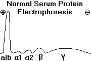
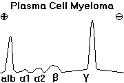
The monoclonal protein (immunoglobulin or chain) produced by an abnormal clone of
multiple
myeloma cells is called the M-PROTEIN.
If there's a complete antibody, you'll see it on serum protein electrophoresis.
Free light chains may be produced along with, or instead of, a complete immunoglobulin. They pass
easily through the glomerular basement membrane, so you will probably not find them in the
bloodstream if the kidneys are working. Instead, they accumulate in the urine, where they are called
BENCE-JONES PROTEIN. About 2/3 of myeloma patients produce Bence-Jones protein.
Later on, Bence-Jones protein plugs up the renal tubules, and contributes to "myeloma kidney", which
we'll study in the "renal pathology" section.
You remember that plasma cell myeloma is an important cause of amyloidosis AL,
which doesn't
help renal function, either.
Here's a breakdown on types of M-proteins:
55%...IgG
25%...IgA
1%... IgE, IgD, or IgM monomer
18%... Bence-Jones protein only
1%... no M-protein.
A PARAPROTEIN is an abundant, useless, monoclonal protein in the bloodstream. All M-proteins are
paraproteins; you'll meet others. Lots of an M-protein will produce rouleaux formation; we'll talk more
about this in "Clinical Pathology".
NOTE: As a rule, plasma cell myeloma does not make IgM pentamers. Waldenstrom's does this, and
you won't see the typical bone changes.
To make the diagnosis, you will want to find an overabundance (>15% or so) or sheets of plasma cells
(typical or weird-looking) on bone marrow.
{16554} plasma cell myeloma, cells
{16556} plasma cell myeloma, cells
{13772} plasma cell myeloma, marrow aspirate
{27330} plasma cell myeloma, marrow aspirate
{13775} plasma cell myeloma, bone marrow section
{10751} * "grape cell"
{42054} * "flame cell" (named for its staining properties)
Normally, only around 3% of bone marrow cells are plasma cells, but whenever there is widespread B-cell activation, their
number can increase substantially.
* Future pathologists: In reactive plasmacytic disorders, plasma cells encircle the vessels. In plasma cell
myeloma, you'll probably find plasma cells encircling fat cells.
Patients are now often getting apparent cures.
Paraneoplastic problems are the greatest problem in plasma cell myeloma.
Be alert for:
- osteoporosis
- pathologic fractures
- hypercalcemia (several mechanisms)
- infections (myeloma cells suppress normal plasma cells)
- anemia and neutropenia (crowding out of normal cells)
- kidney failure (precipitate and/or amyloid)
- * myeloma neuropathy (infiltration, compression, vincristine)
- amyloidosis B (10% of myeloma patients; cardiac problems)
- plasma cell leukemia (a terminal stage)
{17273} myeloma kidney, Bence-Jones casts with foreign body reaction
{17274} myeloma kidney, Bence-Jones casts with foreign body reaction
The tumor generally excites no fibrous or osteoblastic response. At autopsy, the tumor masses (if
distinguishable) look and feel like reddish-gray jelly.
Prognosis is much better, nowadays due both to chemotherapy and to bisphosphonate management
of bone disease. The ongoing "total therapy" studies
is reporting prolonged remissions (cures?) in many patients (updates Blood 112:
3115, 2008; Cancer 133: 355, 2008; Cancer 112: 2720, 2008).
The monoclonal bortezomib (proteasome inhibitor) is very promising (updates Cancer 110:
1042, 2007; Cancer 112: 1529, 2008).
Thalidomide for refractory
myeloma NEJM 341: 1565, 1999.
This is now mainstream.
The disease often simply smolders, and if there are no symptoms,
perhaps it's best just to give bisphosphonates prophylactically (Cancer 113: 1588, 2008).
OTHER PLASMA-CELL PROBLEMS ("plasma cell dyscrasias", an archaic term)
There are a variety of other MONOCLONAL PLASMA CELL PROLIFERATIONS.
We already looked at WALDENSTROM'S MACROGLOBULINEMIA and the
HEAVY-CHAIN DISEASES under "non-Hodgkin's
lymphomas". These are cancers of small lymphocytes with "plasmacytoid" features.
SOLITARY PLASMACYTOMAS may appear benign grossly and microscopically, and they may or may not
produce immunoglobulins.
Those in bone almost always recur as plasma cell myeloma.
Those in extra-osseous sites ("plasmacytic lymphoma") may often be resected for cure.

MONOCLONAL GAMMOPATHY OF UNCERTAIN SIGNIFICANCE ("MGUS", the old "benign monoclonal
gammopathy") affects maybe 3-5% of older adults (prevalence NEJM 354: 1362, 2006;
old texts are wrong to suggest it is less
common than plasma cell myeloma).
This is best considered a benign, disseminated proliferation of plasma cells with some potential to
transform into malignancy.
The tumor cells produce a single, complete immunoglobulin
(usually IgG) that may be detected on serum protein
electrophoresis.
Maybe 1/4 of these people eventually go on to get sick from plasma cell myeloma, amyloidosis AL, or
macroglobulinemia (Mayo Clin. Proc. 68: 26, 1993); newer work gives the rate
at about 1%/year (NEJM 346: 564, 2002)
AMYLOIDOSIS B may arise in this setting, and probably all non-cancer-related amyloidosis AL cases have
a hyperactive clone of plasma cells.
Smoldering myeloma (NEJM 356: 2582, 2007) features 10% or more
plasma cells in the marrow, and an M-protein of myeloma proportions, but no
signs of end-organ damage. It often, but not always, proceeds to myeloma over the years.
 MGUS
MGUS
Pittsburgh Pathology Cases
* POEMS may arise: polyneuropathy, organomegaly, endocrinopathy (thyroid/gonads), monoclonal
gammopathy (usually IgA-lambda), and skin changes.
The molecular etiology remains elusive
(Am. J. Resp. Crit. Care Med. 157: 907, 1998).
* CRYOGLOBULINEMIA TYPE I is a monoclonal immunoglobulin of marginal solubility. More about this in
"Clinical Pathology"!
Follow these people up for decades, and around one in four will get some kind of serious gammopathy
(Mayo Clin. Proc. 68: 26, 1993).

POLYCLONAL ACTIVATION OF PLASMA CELLS ("polyclonal gammopathy") is a common finding in clinical
medicine. Situations worth remembering:
- really bad, long-term infections
- lupus, rheumatoid arthritis, other "autoimmune"
- liver disease
- AIDS
THE LANGERHANS CELL HISTIOCYTOSIS FAMILY ("LCH", "Histiocytosis X", "disseminated
histiocytosis"; * R&F "differentiated histiocytosis" is a typo); review for clinicians
J. Ped. 127: 1, 1995;
Cancer 85: 2278, 1999.
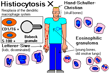
A group (probably a continuum) of lesions that are probably honest-to-goodness tumors of
Langerhans-type histiocytes, a class of dendritic macrophages.
Langerhans cells in health and disease are characterized by intracellular
BIRBECK GRANULES ("histiocytosis
X bodies"), pentalaminar tennis-racket shaped structures of unknown significance.
* Langerin, a lectin specific to stimulated
Langerhans cells: Immunity 12: 71, 2000.
{09095} Birbeck granules
{09097} Birbeck granules
 Histiocytosis X
Pittsburgh Illustrated Case
 Histiocytosis X with Birbeck granules
Lung pathology series
Dr. Warnock's Collection
 Eosinophilic granuloma of the lung
Lung pathology series
Dr. Warnock's Collection
In tissue, you will probably see a range of cells from "blasts" to well-differentiated Langerhans cells.
The former claim that histiocytosis X is "polyclonal" probably resulted from confusion of the tumor cells
with non-neoplastic inflammatory cells that had entered the tumor.
By the mid-1990's we knew
the disease was clonal, hence a real neoplasm
(NEJM 331: 154, 1994; Br. Med. J. 310: 74, 1995; Lancet 344: 1717, 1994).
Future pathologists: Histiocytosis X and the
dendritic macrophages from which it derives stain for CD1/T6. They also stain with S-100.
The old names are passing out of use, but you might perhaps see the syndromes:
LETTERER-SIWE DISEASE ("acute disseminated histiocytosis", "multifocal multisystem LCH") affects small children and involves most of
the body's organs. These children are now often cured with elaborate chemotherapy.
{23392} Letterer-Siwe disease. Weird histiocytes ("coffee-bean nuclei, even"). Trust me.
{13688} eosinophilic granuloma
{13691} eosinophilic granuloma
{09043} eosinophilic granuloma, EM, coffee-bean nucleus (left) and eosinophil (right)
HAND-SCHÜLLER-CHRISTIAN DISEASE ("multifocal unisystem LCH", affects the skull bones and perhaps -- look for diabetes
insipidus, proptosis, lytic skull lesions, fever, and rash. It's intermediate between
the other two in severity.
And not in the classic scheme, but recognized now thanks to improved
pathology techniques, CUTANEOUS LANGERHANS CELL HISTIOCYTOSIS, a disease
of infants that often self-cures (Arch. Derm. 146: 149, 2010).
{10481} Hand-Schüller-Christian disease. Weird histiocytes. Trust me.
{21779} skull in Hand-Schüller-Christian disease
* CHESTER-ERDHEIM DISEASE ("lipid granulomatosis";
"cholesterol granulomatosis") is a rare illness in which lipid-laden
non-dendritic-type macrophages infiltrate the tissues. Thankfully
rare, it is clonal and seems to be a neoplasm (Hum. Path. 30: 1093, 1999).
THE SPLEEN AND ITS PROBLEMS
* Every man has his own ways of courting
the female sex. I should not, myself, choose to do it with photographs
of spleens, diseased or otherwise. -- Agatha
Christie, "The Moving Finger"
The healthy spleen weighs 50-250 gm or less. You remember that the cells
right around the arteries in the white pulp are T-cells, that there are likely to be B-cell nodules,
and that the Littoral cells lining the sinuses express both macrophage and endothelial markers.
The spleen almost never gets biopsied, as it is so likely to rupture.
SPLENOMEGALY must be substantial (800 gm or so) to be palpable. Causes worth remembering:
INFECTIONS
Malaria (huge spleens)
Infectious mononucleosis family (see above)
Bacterial endocarditis (don't miss this one)
Most other bad infections (NOTE: A "septic spleen" feels soft, unlike most of the other big spleens)
CONGESTION (if longstanding, becomes "fibrocongestive")
DISEASES OF WHITE CELLS
Primary myelofibrosis (huge spleens)
SPLENIC OVER-DESTRUCTION OF BLOOD CELLS
Hereditary spherocytosis
Hemoglobinopathies and bad thalassemia
Immune hemolytic anemia
Immune thrombocytopenic purpura
IMMUNOREACTIVE HYPERPLASIA
Lupus
Rheumatoid arthritis
Graft rejection
STORAGE DISEASES (huge spleen)
Gaucher's (very big, wadded-kleenex macrophages)
Niemann-Pick's (very big, foamy macrophages)
Hunter's
Hurler's
SARCOIDOSIS
AMYLOIDOSIS (sago, lard)
HYPERSPLENISM is said to be present when an enlarged spleen destroys normal formed elements of blood too
readily. The three causes you'll probably see are: (1) cirrhosis; (2) rheumatoid arthritis (the
serious "Felty's syndrome"), and (3) Gaucher's disease. It's also one cause of thrombocytopenia
in some leukemia and lymphoma patients.
{00239} Gaucher's disease, spleen
{09864} Gaucher's disease, spleen
{16216} Gaucher's disease, watered-silk ("wadded kleenex") cell from spleen
ACCESSORY SPLEENS (one or more) are present somewhere in the abdomen in about 25% of autopsies. If
you need a splenectomy for a medical disease (i.e., immune thrombocytopenic purpura, hereditary
spherocytosis), you must hope that your surgeon does not overlook a large accessory spleen.
SEPTIC SPLEEN ("nonspecific acute splenitis") is typical of serious bacterial infections. Loaded with polys
and abnormally soft, the old gourmet pathologists made the comparison to "tomato paste",
which is very much resembles.
Exactly why the spleen becomes like this in deaths from sepsis,
and never anything else, remains as mysterious as sepsis itself.
You'll see profound loss of the B-cells and T-helper cells
around the white pulp,
and apoptosis of the dendritic reticular cells that maintain the structure
of the spleen (NEJM 348: 138, 2003).
HYPERPLASTIC SPLEEN usually means large germinal follicles in the white pulp. Think of systemic
autoimmune disease, infectious mononucleosis, graft rejection, etc., etc.
In INFECTIOUS MONONUCLEOSIS, the spleen also becomes infiltrated with activated T-cells that give a
malignant appearance. The capsule is stretched and infiltrated, making it more fragile. You're unlikely to see such a spleen unless it is removed
because of rupture (sports, overzealous physical exam).
* Future pathologists: Telling hyperplasias from lymphomas in the spleen is one of your toughest calls.
For help, see Am. J. Clin. Path. 99: 486, 1993.
INFARCTS are common in the spleen, and may result from atheroembolization (the twisty splenic artery
is the most severely affected in the body), left-sided endocarditis, or infiltrative disease.
* Necrosis in a blood-bloated spleen (typically, in sicklers) is likely to produce iron- and calcium-rich
scars called GAMNA-GANDY BODIES. The "autosplenectomized" spleen of an older sickle-cell disease
patient is mostly composed of such scars.
 Sickle cell disease
Autosplenectomy
WebPath Photo
PRIMARY NEOPLASMS of the spleen are uncommon. Benign tumors are almost never of any importance.
Any lymphoma or endothelial neoplasm can arise here. METASTASES to the spleen are expected in most
leukemias and Hodgkin's and non-Hodgkin's lymphomas, but carcinomas and sarcomas very seldom
grow in the spleen.
RUPTURED SPLEEN results from blows (hard if you're healthy, lighter blows suffice for those with infectious
mononucleosis; remember CPR as a cause Br. Med. J. 322: 480, 2001).
Intraperitoneal hemorrhage results in a trip to surgery. If a lot of pulp escapes into the
peritoneal cavity, the patient may heal with hundreds of mini-spleens over the peritoneal cavity
(SPLENOSIS).
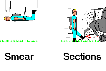
You remember
the difference between sections and smears, right?

* SLICE OF LIFE REVIEW: BLOOD CELLS
10110 ff blood
{10766} leukemia, hairy cell and normal
{12275} anemia, iron deficiency; normal
{13715} lymphocyte, normal
{13868} red blood cell, normal blood
{13910} red blood cell, normal
{14702} polymorphonuclear leukocyte, normal
{14703} polymorphonuclear leukocyte, normal
{14704} polymorphonuclear leukocyte, normal
{14705} polymorphonuclear leukocyte, normal
{14705} polymorphonuclear leukocyte, normal
{14706} polymorphonuclear leukocyte, normal
{14707} polymorphonuclear leukocyte, normal
{14708} eosinophil, normal
{14709} eosinophil, normal
{14710} basophil, normal
{14711} basophil, normal
{14712} monocyte, normal
{14713} monocyte, normal
{14714} monocyte, normal
{14715} monocyte, normal
{14716} lymphocyte, large
{14717} lymphocyte, large
{14718} lymphocyte, normal
{14719} lymphocyte, normal
{14720} lymphocyte, normal
{14721} lymphocyte, normal
{14722} reticulocytes, normal
{14723} reticulocytes, normal
{14724} red blood cell, abnormal
{14725} red blood cell, abnormal
{14726} platelets, normal
{14727} platelets, normal
{14728} pronormoblast, normal
{14729} pronormoblast, normal
{14730} basophilic normoblast, normal
{14731} basophilic normoblast, normal
{14732} normoblast
{14733} normoblast
{14734} polymorphonuclear leukocyte & * lymphocyte
{14735} polymorphonuclear leukocyte & * lymphocyte
{14736} normoblast series
{14737} normoblast series labelled
{14738} myelocyte, normal
{14739} myelocyte, normal
{14740} * granulocyte series
{14741} * granulocyte series (labelled)
{14742} myelocyte, band form
{14743} myelocyte, band form
{14744} myelocyte, normal
{14745} myelocyte, normal
{14746} myelocyte, normal
{14747} myelocyte, normal
{14748} myelocyte, normal
{14749} myelocyte, normal
{14750} myelocyte, normal
{14751} myelocyte & megakaryocyte, normal
{14752} myelocyte & megakaryocyte, normal
{15193} plasma cell, #23
{15205} thymus, adult
{15564} thymus, normal
{15565} thymus, normal
{15566} thymus, normal
{15567} thymus, normal
{16175} red blood cell, normal
{20782} polymorphonuclear leukocyte, normal
{20783} monocyte
{20784} platelets, circulating blood
{20785} monocyte
{26230} polymorphonuclear leukocyte, normal
{40179} thymus, normal
{46538} red cell, normal
* SLICE OF LIFE REVIEW: LYMPHOID ORGANS
{11750} spleen, normal
{11751} spleen, normal
{11753} lymph node, normal
{11797} spleen, normal
{11805} spleen, normal unfixed
{14753} thymus, human fetal
{14754} thymus, human fetal
{14755} thymus, juvenile
{14756} thymus, juvenile
{14757} thymus, adult
{14758} thymus, adult
{14759} thymus, juvenile
{14760} thymus, juvenile
{14761} hassall's corpuscles
{14762} hassall's corpuscles
{14763} hassall's corpuscles
{14764} hassall's corpuscles
{14765} thymus (septum)
{14766} thymus (septum)
{14767} spleen, normal
{14768} spleen, normal
{14769} spleen, pulp
{14770} spleen, pulp
{14771} spleen (trabeculae), normal
{14772} spleen (trabeculae), normal
{14773} spleen (trabecular artery), normal
{14774} spleen (germinal center), normal
{14775} spleen (germinal center), normal
{14776} spleen (venous sinus), normal
{14777} spleen (venous sinus), normal
{14778} spleen (scanning em)
{14779} spleen (scanning em)
{14780} lymph node, normal
{14781} lymph node, normal
{14782} lymph node cortex, normal
{14783} lymph node cortex, normal
{14784} lymph node, medulla
{14785} lymph node, medulla
{14786} lymph node, normal
{14787} lymph node, normal
{15189} lymph node and subcapsular sinus, #23
{15190} lymph node, primary nodule
{15191} lymph node, germinal center
{15192} lymph node, medulla
{15194} spleen, #24
{15195} spleen, * red pulp and white pulp
{15196} spleen, central artery
{15197} spleen, central artery and germinal cent
{15198} spleen, trabeculae
{15199} thymus, #25
{15200} thymus, cortex
{15201} thymus, hassall's corpuscle
{15202} thymus, medulla
{15203} thymus, epithelial reticular cell
{15568} spleen, normal
{15569} spleen, normal
{15570} spleen, normal
{15571} spleen, normal
{15769} spleen, normal
{15770} spleen, normal
{20200} spleen, normal
{20799} lymph node, overview
{20800} lymph node, cortex
{20801} lymph node, medulla
{20802} lymph node, subcapsular sinus
{20803} lymph node, secondary nodule
{20804} lymph node, primary nodule
{20805} spleen, normal histology
{20806} spleen, red pulp
{20807} spleen, white pulp
{20808} spleen, central artery
{20809} spleen, red pulp
{20810} spleen, secondary nodule
{20811} spleen, trabecula
{20812} thymus, overview
{20813} thymus, medulla
{20814} thymus, cortex
{20815} thymus, hassall's corpuscle
{20827} tonsil, palatine
{20828} tonsil, pharyngeal
{24782} lymph node, normal
{24783} lymph node, normal
{36344} lymph node, normal
{36347} lymph node, normal
{36350} lymph node, normal cytology
{36353} lymph node, normal cytology


 Waldenstrom's
 Post-Transplant Neoplasia


