 NORMAL ANATOMY
Title: Tumors, Aneurysms, Vasculitis
Date & Time: Friday, February 8, 2013 at 11 AM
Lecturer: The Pathology Team
QUIZBANK: Hemodynamic #'s 1-30, Vessels (all)
The term "arteriosclerosis" (literally, "hardening of the arteries") should be avoided by physicians. It includes (1) atherosclerosis; (2) Monckeberg's medial calcific sclerosis; and (3) arteriolar sclerosis (hyaline, hyperplastic, intimal fibrosis). Unqualified, the term usually means "atherosclerosis".
The vascular intima looks simple but isn't. Endothelial cells must maintain their no-stick inner surfaces, help constrict and dilate vessels, and heal damaged vessels. Myointimal cells and macrophages, located between the endothelium and the internal elastic membrane, are the principal actors in atherosclerosis.
Atherosclerosis is a stereotyped response of the inner surfaces of large arteries to a variety of insults. In this disease, the cells between the endothelium and the internal elastic membrane take up cholesterol-rich lipid, which then causes harm. Lesions progress from FATTY STREAKS to FIBROUS PLAQUES to COMPLICATED FIBROUS PLAQUES; they can also regress.
Atherosclerosis may calcify, but the problem in atherosclerosis is not the dystrophic calcification. Monckeberg's medial calcific sclerosis just means dystrophic calcification of the media of an artery, but it is almost never a real problem.
Atherosclerosis is an ancient disease, present in some Egyptian mummies (JAMA 302: 2091, 2009 -- the lesions are real but look quite mild by today's standards).
Atherosclerosis was THE great killer of 20th century North Americans. The epidemic peaked in 1968, and since then the decline has been spectacular, due more than anything else to healthier lifestyles (JAMA 277: 535, 1997; it's been steady since the mid-1980's: NEJM 339: 861, 1998). We are beginning to understand how the common risk factors relate to its pathogenesis. Americans are taking steps to protect themselves, and lifestyle changes can almost certainly reverse much of the damage in all but the most advanced lesions.
Hyaline arteriolar sclerosis results from damage to arterioles usually from increased pressure or increased blood glucose. Hyperplastic arteriolar sclerosis involves hyperplasia of the intimal cells; it results from processes that do severe, acute damage to the endothelium. Fibrosis of the intima results from high blood pressure or "just getting older."
NORMAL ANATOMY
ARTERIES carry blood from the heart.
ELASTIC ARTERIES ("large arteries") include the aorta and at least the beginnings of its largest branches. These arteries both propel and dampen the pulse wave. These are distinguished by a preponderance of elastic fibers in their media. The subendothelium of their intimal layers thickens over the course of life through the accumulation of collagen fibers and myointimal cells. The elastic tissue proliferates here. You already know that all elastic tissue slowly breaks down as we age. In older adults, the elastic is largely replaced by collagen. This also results in lengthening and thus tortuosity seen in older people. The adventitia and outer media are nourished by vasa vasora. All arteries depend on the blood within their lumens to nourish their intima and inner media.
MUSCULAR ARTERIES ("medium-sized arteries", "distributing arteries") exhibit smooth muscle in their walls, and may expand and contract to regulate the caliber of the lumen and thus the flow of blood. The intima is similar to that of the elastic arteries, and it thickens similarly. Smooth muscle may pass into the intima through fenestrae in the internal elastic membrane. These fenestrae may become wide in old age and be mistaken for damage from previous vasculitis. The media is bounded on either side by an inner and outer elastic membrane.
SMALL ARTERIES are the major site of autonomic regulation of blood flow, and take the worst beating in hypertension. A rule of thumb is that the wall and lumen should have the same thickness. Thickening of the intima occurs here as well. In sites of inflammation or tumor, it may be quite impressive (* "Friedlander's endarteritis obliterans", discovered by the real Dr. Friedlander, 19th century pathologist Carl, 1847-1887). Hyaline arteriolar sclerosis becomes a problem as we age, especially if we get diabetes or hypertension. There's no outer elastic membrane, and the layers become progressively less distinct as the arteries get smaller.
ARTERIOLES continue the anatomy of the small arteries. Two definitions that have been offered: (1) Arterioles have five or fewer layers of smooth muscle; (2) Arterioles have total diameter 100 microns or less.
VEINS and LYMPHATICS have histologic features that you know. In disease, veins do not usually show so much intimal proliferation and fibrosis as do arteries. The muscle in the wall of a vein is thinner, and in the larger veins tends to be less organized. Very large veins have some layers of elastic outside their muscular layer. Lymphatics run very close to arteries (even closer than the veins), and tend to be small and to have thinner walls than the vein that runs with that artery. It's not always possible to tell lymphatics from veins; if the vessel contains red cells, it's most likely a vein.
![]() Lymphatic vessels in edema
Lymphatic vessels in edema
They are not themselves plugged,
but are dilated from carring away fluid.
ENDOTHELIUM is special stuff. It is permeable to water and the small inorganic ions. It transports a little bit of blood protein by pinocytosis. Electron microscopists recognize it by the WEIBEL-PALADE BODIES (puh-LAH-dee, made of von Willebrand's factor). It can contract, to regulate capillary flow. It produces some of the subendothelial connective tissue. It also makes substances: (1) Prostacyclin (to keep its surface slippery); (2) Von Willebrand's factor; (3) Endothelin (a vasoconstrictor peptide); (4) Endothelial-derived relaxation factor (nitric oxide, EDRF; see Nature 368: 62, 1994).
VASCULAR SMOOTH MUSCLE is also special. It has LDL receptors. It can get into the intima through holes in the internal elastic membrane. Both facts will become important when we study atherogenesis.
 BIRTH DEFECTS INVOLVING VESSELS
There are many variations on the normal anatomy of arteries.
Malformations of the coronary arteries may first announce themselves by causing sudden death. More about this soon.
The familiar red "birthmarks" are hemangiomas, and will be covered with "tumors".
The only other birth defects worth mentioning are BERRY ANEURYSMS and ARTERIOVENOUS MALFORMATIONS ("AV malformations", "AV fistulas", "AV aneurysms", etc.)
AV malformations involve a tangle of abnormal medium-sized vessels connecting a large artery and a large vein. The problem is shunting of the blood away from the territory that should be supplied by the artery. The vein will tend to expand ("aneurysm").
Sometimes the AV malformation is a mass of wormy vessels ("cirsoid aneurysm", "racemose aneurysm"; apparently endemic among Klingons). This is most common in the brain, where subarachnoid hemorrhage is the dread complication.
* Future pathologists: It's good to be able to distinguish a baby's AV malformation (which won't go away) from a baby's hemangioma (which probably will go away). An AV malformation does not stain for WT1; a hemangioma that will involute does stain for WT1 (Arch. Derm. 141: 1297, 2005).
* Genetic syndromes that affect vessels include Ehlers-Danlos type IV (the arterial walls become very weak and fragment), neurofibromatosis type I (lack of neurofibromin can cause aneurysms and malformations of the vessels especially in the head, or an artery may simply burst), and Fabry's (storage in the endothelium.)
MONCKEBERG'S MEDIAL CALCIFIC SCLEROSIS
|
|
Dystrophic calcification (sometimes even ossification) of the media of arteries, typically in older adults.
This is a common, banal, pretty much harmless process. At worst, it can widen one's pulse pressure due to decreased aortic compliance, or make "radial artery blood-gas sticks" difficult and hazardous. It may be visible on x-ray.
Despite the term "medial", the internal elastic membrane is always calcified (settled Arch. Path. Lab. Med. 132: 47, 2008).
* Evidently the smooth muscle cells produce at least four proteins that indicate they want to make bone. You can read about it in Circ. 100: 2168, 1999.
ARTERIOLAR SCLEROSIS ("arteriolosclerosis")
|
|
|
Three processes that narrow the lumens of the small arteries and arterioles in some or all of the body
INTIMAL FIBROSIS or (better) "fibroelastic hyperplasia" (and some call it "intimal sclerosis"), is the slow buildup of fibrous tissue (usually with some layers of elastic) in the intima of a small artery. It's a part of aging, and is exacerbated by high blood pressure. It involves smaller arteries (rather than the larger ones, as in atherosclerosis) and doesn't feature the lipid buildup. It also spares the arterioles. The most familiar form is the damage to the small arteries of the kidney seen in longstanding high blood pressure.
HYALINE ARTERIOLAR SCLEROSIS: slow buildup of basement-membrane type material, eventually obliterating the cellular structure of the wall and narrowing the lumen. Mostly the arterioles are involved.
The most common place where we see this is in the arterioles of the kidney in high blood pressure and diabetes.
However, it turns up often, and can appear anywhere. The most familiar causes are
No one really knows why any of these causes "hyaline arteriolosclerosis", but the anatomic pathology is impressive. Hypertension is the least potent. The effects on tissue perfusion, and probably on the sympathetic regulation of blood pressure, are not salutary.
Future pathologists: Look in the fat just outside the adrenal capsules to get a good idea about the extent of systemic hyaline arteriolosclerosis. Lots of hyaline change of the small arteries here is a great marker for longstanding hypertension. "Binswanger's encephalopathy" is a dread, Alzheimer-like dementia caused by hyaline arteriolosclerosis of the brain.
* If you like, you can consider amyloidosis of small arteries, which hyalinizes but is less likely to cause much stenosis, to be another cause of hyaline arteriolar sclerosis. No one will argue or care.
{11777} hyaline arteriolar sclerosis
{40267} hyaline arteriolar sclerosis
{40347} hyaline arteriolar sclerosis

HYPERPLASTIC ARTERIOLAR SCLEROSIS: Concentric, often rapid proliferation of the intimal (or sometimes smooth muscle) cells of an arteriole. "Onion-skin arteriole".
Fortunately uncommon. The causes are
{39559} hyperplastic arteriolar sclerosis in pulmonary hypertension
{24854} hyperplastic arteriolar sclerosis, scleroderma kidney
|
|
Note that all these are processes that specifically damage the intima of vessels, which presumably undergo hyperplasia in response. This is a much more aggressive process. (You could think of it as a callus of the endothelium.)
Note that any of these processes can be severe enough to cause some necrosis of the vessel. Malignant hypertension is usually this severe.
Don't confuse either of these processes with the inexorable FIBROUS THICKENING OF THE INTIMA OF SMALL ARTERIES in mild hypertension and normal aging. This is so common and so important that IT DOESN'T HAVE A NAME BEYOND "INTIMAL FIBROSIS".
TRANSPLANT VASCULOPATHY is a concentric fibrous thickening, mostly confined to the intima, in allografts (heart, kidney, liver, others) that have survived for a long time; the process develops rather abruptly, and while immunity must be a factor, it cannot be the only explanation. See Am. Heart. J. 129: 791, 1995.
THE VASCULITIS FAMILY (update Am. J. Clin. Path. 124(S): S-84, 2005)
|
|
|
Scarring can narrow a vessel, or cause an artery to balloon (aneurysm).
An inflamed may rupture (petechiae, purpura).
POLYARTERITIS NODOSA
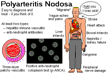
Due to hepatitis B infection (antigen-antibody complexes), anti-myeloperoxidase disease ("anti-MPO", "anti-neutrophil cytoplasmic antibody disease", now distinguished from "true polyarteritis nodosa"), or "idiopathic".
The principal discussion of polyarteritis nodosa, Wegener's, and their family comes under immunopathology. Remember classic polyarteritis nodosa as an important cause of infarcts anywhere in the body except lung.
LEUKOCYTOCLASTIC VASCULITIS
Generally type III immune injury of the venules, often diagnosed on skin biopsy (the patient has "palpable purpura"). Common, but infarcts and serious damage are fortunately rare.
"Leukocytoclastic" refers to the dead neutrophils lying about, visible as nuclear dust.
{14284} leukocytoclastic vasculitis
{14286} leukocytoclastic vasculitis
{14287} leukocytoclastic vasculitis
{14289} leukocytoclastic vasculitis
{14290} leukocytoclastic vasculitis
{14292} leukocytoclastic vasculitis
{14293} leukocytoclastic vasculitis
{14294} leukocytoclastic vasculitis
{14295} leukocytoclastic vasculitis
{14296} leukocytoclastic vasculitis
{14298} leukocytoclastic vasculitis
Mostly this results from taking medicines. Less common causes are cryoglobulinemia (how?), lupus and its kindred, and the antigenemia of HBV and malignancy.
WEGENER'S GRANULOMATOSIS
A vasculitis, usually with granulomas, caused by an anti-proteinase 3 autoantibody (anti-PR3). Lung cavities, segmental necrotizing glomerulonephritis with crescents, and/or vanishing nose, all most likely with granulomas. We'll talk more about this in "immuno".
CHURG-STRAUSS DISEASE
{38497} old burned-out Wegener's, without good granulomas
TEMPORAL ARTERITIS ("cranial giant cell arteritis", when elsewhere "giant cell arteritis")
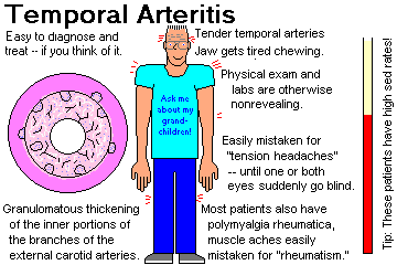
A disease of older folks, mostly over 60 (nobody's immune), still of unknown etiology, in which the macrophages seem to become angry with the internal elastic membrane of the arteries of the external carotid system.
"Jaw claudication" (tired jaw on chewing) is a picturesque syndrome, but sudden blindness (the dread complication) is a catastrophe. Despite the conventional wisdom that the vertebrobasilar and internal carotid systems are not involved much, one group found that around 3% of biopsy-proven "temporal arteritis" patients had a stoke between the onset of symptoms and the initiation of glucocorticoid treatment (Medicine 88: 227, 2009).
Many of these patients also suffer from pain and weakness in the muscles, the distinctive POLYMYALGIA RHEUMATICA (Lancet 381: 63, 2013). Future clinicians: The diagnosis is supported by finding everything normal on physical exam except perhaps for tender temporal arteries, plus normal labs except for a high "sed rate".
Only recently has it become clear that there is systemic overproduction of interleukins 1 and 6 for some reason, and activation of macrophages in the vascular intima. There is still a great deal that' s unknown (Ann. Int. Med. 139: 505, 2003).
Some folks will treat just on the clinical findings. Or prove your diagnosis with a temporal artery biopsy, which may or may not show granulomas on the inner elastic membrane. There is also a striking non-granulomatous fibrous proliferation of the intima. When in doubt, treat with prednisone or some similar glucocorticoid.
* On ultrasound, the aortic arch and its vessels are often thick as well (Rheumatology 51: 730, 2012).
* A histologic variant spares the intima but involves the vasa vasora / small vessels near the artery. Artifact of sampling, or separate entity, it's probably best to consider this temporal arteritis as well (Arth. Rheum. 64: 549, 2012). * Methotrexate to supplement the prednisone for a more-effective, more-tolerable treatment: Arth. Rheum. 56: 2789, 2007.
* The British think the temporal artery biopsy should be 1 cm long (Br. J. Surg. 98: 1556, 2011).
* A variant involves the female genital tract (South. Med. J. 98: 469, 2005).
* How long to treat: Arch. Int. Med. 159: 577, 1999.
Polymyalgia rheumatica can occur in young people and/or in the absence of elevated sed rate (Arch. Int. Med. 157S: 317, 1997). When in doubt, treat.
* Future pathologists: Biopsies remain positive even after several weeks of glucocorticoid therapy (Br. J. Ophth. 86: 530, 2002).
{22095} temporal arteritis
{22096} temporal arteritis
{22098} temporal arteritis
{24777} temporal arteritis
{28019} temporal arteritis
TAKAYASU'S PULSELESS DISEASE ("aortic arch disease", etc.)
A fortunately-rare, idiopathic disease of younger adults (almost always) in which the aortic arch and its great branches thicken and their ostia become stenotic, strangling off blood flow to the upper part of the body.
No one knows the cause, and the histology is nonspecific, with granulomas, giant cells, lymphocytes, plasma cells, and so forth, in addition to the fibrosis and contraction.
* The molecular biology of temporal arteritis and Takayasu's is evidently similar. What's known: NEJM 349: 160, 2003. More on Takayasu's and temporal arteritis as a continuum: Medicine 88: 183 & 221, 2009. Watch for aspirin to be added to the glucocorticoid regimens, to prevent platelet-related intimal fibrosis in both diseases.
Whatever the real cause, surgical repair of the involved arch and branches is now giving excellent results (Ann. Thor. Surg. 81: 178, 2006). On medical therapy, or when surgery is not possible, the long-term prognosis is generally not good (Arth. Rheum. 56: 1000, 2007).
{48983} Takayasu's
COGAN'S DISEASE is another thankfully-rare disease usually affecting young adults. It features abrupt onset of nerve deafness, interstitial keratitis (you'll see it only on a slit lamp exam), and/or a systemic vasculitis often with aortic aneurysm formation. It's evidently caused by an autoantibody against inner ear and endothelium (Lancet 360: 915, 2002).
KAWASAKI'S DISEASE ("mucocutaneous lymph node syndrome"; Ped. Clin. N.A. 46: 313, 1999; Am. Fam. Phys. 59: 3093, 1999; Lancet 364: 533, 2004; Heart 95: 787, 2009)
A febrile disease that resembles adult polyarteritis nodosa histologically but occurs in babies and toddlers, mostly of Japanese or Korean ancestry (no matter where they live now). The larger arteries get the most severe involvement.
The fact that almost all patients are around 2-5 years, the fact that occasionally an older child or adult gets the disease, the fact that there are outbreaks, and the fact that babies don't get it as long as they have maternal antibody all tell me the cause is an unidentified, ubiquitous virus.
You'll want to see five of these six signs:
Most patients are of Japanese of Korean ancestry, regardless of where they live, but no HLA links are found.
The most serious concern is coronary vasculitis, which causes myocardial infarcts. Healing can produce coronary aneurysms, etc. See Arch. Dis. Child. 87: 145, 2002.
* By electron microscopy, the endothelial cells are separated and perforated, rendering them hyperpermeable; again, no one knows why (Circulation 105: 766, 2002).
We treat Kawasaki's with aspirin and intravenous immunoglobulin. The outcome is good unless coronary disease becomes apparent.
Even in youngsters with coronary artery aneurysms, with today's interventions the long-term prognosis is pretty good (Circulation 123: 1836, 2011).
|
BUERGER'S DISEASE ("thromboangiitis obliterans": review
Angiology 47: 419, 1996; update Am. J. Med. Sci. 337: 285, 2009)
A disease of smokers, usually young men, in which the small neurovascular bundles in the extremities become inflamed and undergo thrombosis. No one has a clue as to the real etiology, beyond the link to tobacco smoking. It's also an ultra-rare complication of marijuana smoking (Br. J. Derm. 152: 166, 2005). The typical patient, after losing all his fingers, holds his cigaret between his last two toes. Not a pretty sight. The prognosis is hopeless unless the patient stops smoking. The anatomic pathology remains poorly worked-out. Neutrophils touching giant cells within thrombi is supposed to be characteristic. The most recent study (Virchows Archiv 436: 59, 2000) found instead that an intact internal elastic membrane, fibrosis much worse in the adventitia than anywhere else, endothelial swelling in the vasa vasora, and onionskinning of recanalization vessels were most helpful. |

|
INFLAMMATORY AORTIC ANEURYSM (JAMA 297: 395, 2007)
The overwhelming majority of patients are smokers. Otherwise, the etiology is unknown. I predict that the molecular cause of Buerger's and inflammatory aneurysm will be found at the same time.
Unlike classic atherosclerotic aneurysms, the outer surface is shiny-white with prominent little vessels, and usually there are fibrous adhesions to the nearby structures. The histopathology is intense inflammation and fibrosis of the adventitia.
INFECTIOUS ARTERITIS
Rickettsial disease, syphilis![]() ,
septic emboli (look for "Roth's spots!"), walls of abscesses, and a host of
others.
,
septic emboli (look for "Roth's spots!"), walls of abscesses, and a host of
others.
Worth mentioning here: A MYCOTIC ANEURYSM is a spot at a branch-point of an artery where a septic embolus (usually) has lodged and set up an infection, weakening the wall. ("Mycotic" is an unfortunate misnomer, since fungi aren't usually the culprits.)
Among the fungi, remember aspergillus and mucormycosis as especially good at invading vessels.
RAYNAUD"S DISEASE / PHENOMENON
Spasm and occlusion of the arteries supplying the fingers, which turn white ("pallor"), then red ("suffusion"), then blue ("cyanosis"). Triggered by cold weather, it's most often idiopathic; known causes range from vasculitis syndromes to operating jack-hammers.
Scleroderma patients and some others have this process greatly exacerbated by hyperplastic arteriolar sclerosis in the digital arteries.
If it's a bother, get out the calcium-channel blockers, and/or a nice warm pair of gloves.
{24503} Raynaud's
{25459} Raynaud's
{39657} Raynaud's
{39654} Kawasaki's?
{39655} Kawasaki's?
{48983} Takayasu's
The common pathophysiology is loss of vascular control; they will first become constricted or blocked, then dilate and become inflamed.
The red color of affected body parts probably results from opening of arteriovenous channels and closure of precapillary sphincters. Lack of nutritive blood flow contributes to the pain, while the excess non-nutrative blood flow causes a burning sensation.
Long-mysterious, it's now pretty clear that it's a often a small-fiber neuropathy (Arch. Derm. 139: 1337, 2003; Brain 126: 567, 2003; update Anes. Anal. 104: 438, 2007).
There's a hereditary form with mutated neuronal sodium channels (Neurology 67: 1538, 2006), and it is possible that the adult acquired form results from autoantibodies against this channel (Am. J. Med. Sci. 335: 320, 2008). These patients present a major pain-management problem -- Mayo's has a series (Arch. Derm. 144: 1578, 2009).
Myeloproliferative diseases with extremely high platelet counts also produce erythromelalgia, probably by the platelets plugging vessels.
ATHEROSCLEROTIC AORTIC ANEURYSMS
|
|
|
ATHEROSCLEROSIS often causes aneurysms, usually distally in the aorta, above the level of the iliacs. When the abdominal aortic diameter exceeds 3.0 cm, it's an aneurysm. Exceed 5 cm or so and it's likely to burst (retroperitoneally, intra-peritoneally, intra-duodenal), with (usually) lethal consequences. Patients may complain first of back pain, etc. Aneurysms are always lined by thrombus (why?), which sometimes embolizes. Iliac aneurysms are also common; basilar artery aneurysms seldom rupture but may compress important things.
Occasionally these get infected; the usual bug is salmonella (Am. J. For. Med. Path. 23: 382, 2002).
* Albert Einstein's physicians knew he had an aneurysm, but when it burst, they got focused on his gallbladder instead. He died as a result.
{03665} atherosclerotic aortic aneurysm
{11042} atherosclerotic aortic aneurysm
{11048} atherosclerotic aortic aneurysm, repaired
{11642} atherosclerotic aortic aneurysm
{11645} atherosclerotic aortic aneurysm
{18717} atherosclerotic aortic aneurysm
{20305} atherosclerotic aortic aneurysm
{24780} atherosclerotic aortic aneurysm
{25742} atherosclerotic aortic aneurysm
{04589} atherosclerotic basilar artery aneurysm
{24836} atherosclerotic aneurysm, brain
|
|
![]() Coronary Artery Aneurysm
Coronary Artery Aneurysm
Classic drawing
Adami & McCrae, 1914
SYPHILIS![]() ("lues")
causes ischemic damage (by narrowing / occluding vasa vasora) to the walls of the arteries, and is
famous for causing proximal aneurysms that rupture impressively. Before rupture occurs, look
for the infamous "tree bark" grooves on the intima, as well as occlusion of the coronary and other
ostia and compromise of the aortic valve ring. It's still very much around
(Ann. Thorac. Surg. 92: 1503, 2011).
("lues")
causes ischemic damage (by narrowing / occluding vasa vasora) to the walls of the arteries, and is
famous for causing proximal aneurysms that rupture impressively. Before rupture occurs, look
for the infamous "tree bark" grooves on the intima, as well as occlusion of the coronary and other
ostia and compromise of the aortic valve ring. It's still very much around
(Ann. Thorac. Surg. 92: 1503, 2011).
Historically, we've taught that proximal aortic aneurysms are usually luetic. Nowadays, syphilis is very rare, and atherosclerosis is common. I wouldn't assume an ascending aortic aneurysm is luetic unless the arch is free of significant atherosclerosis.
* "Tree-barking" is just stretch-marks of the aorta. The only reason this is supposed to be "more typical of syphilis than atherosclerosis" is that, in atherosclerosis, the intima is already distorted by the atherosclerotic plaques. I know this, because in my several cases of Marfan-style dilatation of the aortic root, there's always been tree-barking.
{10224} syphilitic aneurysm
{18716} syphilitic aneurysm
![]() Syphilitic aneurysm
Syphilitic aneurysm
Classic patient photo
Adami & McCrae, 1914
|
|
AORTIC DISSECTION
DISSECTING HEMATOMA, often miscalled "dissecting aneurysm", is blood that has entered the wall of the aorta and is following a weak plane ("cystic medial necrosis", actually there are no cysts and no necrosis, just diminished elastic fibers and maybe a little extra mucoid goo).
Think of the blood acting as a chisel under the strokes of the heart.
* A study that questioned whether the elastic is extensively damaged in most people who have experienced acute ascending aortic dissection (Am. J. Card. 108: 1639, 2011; all patients were or had been hypertensive) doesn't square with your lecturer's small series; wait and see on this one.
The lesion often happens over days or longer, and is infamously missed by physicians who didn't listen for aortic regurgitation. People coming to autopsy after death from thoracic aortic dissections have often (1 in 3) visited their physician during the previous week, and generally have very large hearts (from hypertension and/or from aortic regurgitation) -- don't miss it Am. Heart. J. 162: 474, 2011.
![]() Aortic dissection
Aortic dissection
Classic drawing
Adami & McCrae, 1914
![]() Aortic dissection
Aortic dissection
Dr. Heuser
Wikimedia Commons
![]() "Cystic medial necrosis"
"Cystic medial necrosis"
WebPath Photo
This catastrophe results in progressive compromise of arteries, backwards rupture damaging the aortic valve and/or coronary ostia, or further backwards rupture into the pericardial sac or pleural space.
Patients experience a "ripping", agonizing chest pain as the false lumen expands. Michael DeBakey's claim to fame is having classified and devised the surgical treatment for these people. Otherwise, the patient's only hope is to have the blood re-enter the lumen, establishing a "double-barrel aorta", with the false lumen eventually becoming covered with normal endothelium.
Marfan types are more prone to this lesion, but nobody's immune. An epidemic of dissecting aneurysms occurred among turkeys who acquired osteolathyrism after eating beta-NH2-propionitrile in sweet peas.
* The most spectacular multiple aneurysms in medicine are perhaps seen in the Marfan-like Loeys-Dietz syndrome, caused by a mutation in one or the other receptor for transforming growth factor beta (Nat. Genet. 37: 275, 2005).
* Nowadays, if one of your parents or siblings had an aortic dissection, you go get your genes checked; at this writing (2012), seven loci for "familial thoracic aortic aneurysm and dissection" are known.
Minor variants, with limited extension of a bleed into the aortic wall or down around the root, also exist. These may or may not turn into the fully-expressed, deadly disease.
Trauma to the aorta (for example, a car wreck) or another major artery (infamously, the vertebral arteries in a very bad neck manipulation) can cause dissection of blood down a torn media.
"Spontaneous" dissection of arteries in the neck can cause stroke in young people (NEJM 330: 393, 1994).
Aortic dissection resulting from a catheter mishap is well-known, with a risk of about 0.7% (Am. Heart J. 159: 1147, 2010).
* Non-surgical management using a stent-graft: NEJM 340: 1585, 1999 (wow!).
{17467} dissecting aneurysm
{18718} dissecting aneurysm
{20222} dissecting aneurysm
{25747} dissecting aneurysm
|
|
CORONARY ARTERY DISSECTION is thankfully rare. For some reason, it most commonly occurs after childbirth.
VEINS
VARICOSE VEINS
A self-perpetuating process caused by loss of competency in the leg vein valves and support structures.
We lose our elastic fibers as a result of aging, or blow out valves (by getting fat, having babies, straining at stool, standing up doing surgery). The tendency to get varcose veins is hereditary.
The weight of the column of blood doesn't make life easier for the next valve or the support of the next few cm of vein. Get elastic leg wraps and hope for the best. If it gets too hard to pump blood back to the heart by way of the veins, or the weight of the column of blood causes continuous micro-hemorrhages, you will see the familiar hemosiderin STASIS PIGMENTATION and/or ischemic STASIS ULCERS.
Stasis ulcers themselves are a very common, very hard-to-treat problem of older folks. Older explanations ("the poor circulation produces hypoxia") are being supplemented by new work (the weight of blood causes capillaries to rupture, with red cells and plasma proteins being extravasated, with local cytokine activation; Surg. Clin. N.A. 83: 671, 2003.) This fits with there usually being pigmentation here, and with the accompanying local fibrosis (lipofibrosclerosis).
![]() You already know from physiology about ESOPHAGEAL VARICES and
HEMORRHOIDS as evidence of portal
hypertension.
You already know from physiology about ESOPHAGEAL VARICES and
HEMORRHOIDS as evidence of portal
hypertension.
Fortunately, thrombi that may form in varicose veins very seldom embolize.
THROMBOPHLEBITIS ("trombone fleabites", better "phlebothrombosis"; reviews Lancet 353: 479 & 1167, 1999)
Thrombosis of a deep vein, most often in the leg. You're already familiar with the causes, and the dread complication (pulmonary embolus). Usually, pain and swelling result, hence "-itis".
Vicious variants include MILK LEG ("phlegmasia alba / cerulea dolens", thrombosis of the iliac vein, around the time of parturition), BUDD-CHIARI SYNDROME (thrombosis of the hepatic veins) and DURAL SINUS THROMBOSIS.
TROUSSEAU'S MIGRATORY THROMBOPHLEBITIS affects first one vein, then another. The cause is usually cancer of the pancreas, less often another adenocarcinoma.
SUPERIOR VENA CAVA SYNDROME
From occlusion, usually from extrinsic compression by lung cancer.
Patients have dusky skin in the head, upper extremities, and upper chest.
"SVC syndrome" can cause a bad headache just from the markedly increased venous pressure.
INFERIOR VENA CAVA SYNDROME
From occlusion, usually by cancer in para-aortic lymph nodes, or a thrombus-plugged IVC filtre.
Patients have dusky lower bodies and unusual patterns of collateral circulation.
LYMPHATICS
LYMPHANGITIS
Inflammation of lymphatics, usually by a bacterium that's good at invading them (i.e.,
streptococcus![]() group A.) You'll see red streaks running along an extremity, etc.
group A.) You'll see red streaks running along an extremity, etc.
LYMPHEDEMA
Compromise of lymphatic draining of the extremities and/or genitalia. The most important causes are cancer of the cervix, filariasis, iatrogenic (i.e., after the overly-zealous old mastectomies), autosomal dominant ("Milroy's disease", the lymphatics don't form properly), or "idiopathic" (again, sometimes the lymphatics don't form properly). Turner's girls often have lymphedema of the hands and feet.
If there's hyperplasia of the overlying epidermis, it's ELEPHANTIASIS.
"Massive localized lymphedema" looks clinically and microscopically like a sarcoma, but results from lymphatics obstructed by masive obesity (J. Clin. Path. 62: 808, 2009).
![]() Lymphedema
Lymphedema
Patient education site
By a cyberfriend
TUMORS OF BLOOD VESSELS
The cell of origin is the endothelial cell. When the vascular origin of a tumor isn't obvious, we confirm the diagnosis using immunostains (von Willebrand's factor, CD31, CD34); old-timers found Weibel-Palade bodies in the endothelium on electron microscopy.
HEMANGIOMAS
Mostly hamartomas. You'll see a variety of these clinically.
* Future pathologists: There are some rare, exotic tumors of blood vessel origin. Don't worry about them.
CAPILLARY HEMANGIOMAS (little vessels) and CAVERNOUS HEMANGIOMAS (big vessels "resembling erectile tissue") are common on the skin. STORK BITES (backs of the neck and/or forehead of a baby) usually involute (i.e., thrombose and organize) after a few years, while CHERRY ANGIOMAS of the skin start popping up after age 20 or so.
{05888} hemangioma
{12235} hemangioma
{13041} hemangioma
{13042} hemangioma
{13044} hemangioma
{13045} hemangioma
{13047} hemangioma
{13048} hemangioma
{13050} hemangioma
{13051} hemangioma
{13141} hemangioma
{17507} hemangioma in liver
{21832} hemangioma
{21834} hemangioma
{21835} hemangioma
{22116} hemangioma
{22118} hemangioma
{22119} hemangioma
|
|
PORT-WINE STAIN (one form of "nevus flammeus"), fashionable in the Gorbachev era, can be treated by laser if the patient wishes. A hemangioma in the meninges (bad, shunts blood away), generally with an overlying port-wine stain in the V1 area (or check the eyelids; Pediatrics 87: 323, 1991), is STURGE-WEBER syndrome.
{53705} Sturge-Weber
PYOGENIC GRANULOMA (noted misnomer; neither pyogenic nor a granuloma) pops up the gums of pregnant ladies, or the skin of anybody. Grossly, it looks like a rotten cherry and bleeds very easily. Microscopically, it looks like granulation tissue.
{13141} "pyogenic granuloma"
{12786} "pyogenic granuloma"
{12789} "pyogenic granuloma"
{12225} pyogenic granuloma
* EPITHELIOID HEMANGIOMAS are curious bumps of small vessels surrounding a single large one; the connective tissue between the vessels is packed with eosinophils.
* PAPILLARY ENDOTHELIAL HYPERPLASIA is just an unusual way way for a thrombus to organize. Endothelial cells make little papillae. The lack of anaplasia distinguishes it from an angiosarcoma.
Large hemangiomas can consume a person's platelets, the serious KASABACH-MERRITT SYNDROME (Am. J. Clin. Path. 99: 628, 1993; a kid received 6622 units of platelets over 19 months, a new record; Am. J. Med. Sci. 333: 293, 2007 disseminated angiosarcoma).
{12237} Kasabach-Merritt giant hemangioma
KLIPPEL-TRENAUNAY SYNDROME is a huge capillary hemangioma plus enlargement-deformity, always from a somatic mutation (* gene VG5Q: Nature 427: 640, 2004). A single extremity (or perhaps more than one) is involved. Mayo Clin. Proc. 73: 28, 1998.
| * Golfer Casey Martin is the best-known Klippel-Trenaunay patient. In 2001, the US Supreme Court ruled (PGA tours vs. Martin) he could ride in a cart despite the PGA's complaining it would give in an unfair advantage. |
VON HIPPEL-LINDAU anti-oncogene deletion syndrome results from mutated VHL. It features the most troublesome hemangiomas in all of medicine, as well as renal cell carcinoma.
GLOMANGIOMAS (glomus tumors)
Painful tumors of the smooth muscle of the vestigial human GLOMUS organs, little thermoregulatory left-overs from mammalian evolution and found in the fingertips, toes, and coccyx (the rat's tail is the most familiar glomus).
{09017} glomus tumor
TELANGIECTASIS
"Dilatations of the ends of little vessels".
OSLER-WEBER-RENDU TELANGIECTASIAS ("hereditary hemorrhage telangiectasia") result from any of several autosomal dominant genes (* some, but not all, patients lack a binder for transforming growth factor beta). Most do not know they are affected, and simply they think they have "frequent nosebleeds." Patients have little vascular malformations connecting little arteries and little veins along their whole GI tract (often easiest to see on the lips, of course), and often lots of other places. Especially, AV malformations in the lungs (where they tend to be large) can produce right-to-left shunts, which are dangerous. The lesions in the nose and GI tract are prone to bleed. Reviews NEJM 333: 918, 1995; NEJM 345: 325, 2001; clinicians Am. Fam. Phys. 82: 785, 2010.
* Many of these patients also have "idiopathic pulmonary hypertension", also from faulty vascular development; depends on the allele).
The familiar "liver spider" is a centrally dilated artery supplying several little arterioles that blanch when the "spider's body" is pressed.
CANCERS OF THE BLOOD VESSELS
HEMANGIOENDOTHELIOMAS are low-grade cancers of endothelium. They makes little vessels. Leave the diagnosis to the pathologists.
(HEM)ANGIOSARCOMAS (Arch. Path. Lab. Med. 133: 1804, 2009; Ann. Surg. 251: 1098, 2010): A host of aggressive cancers. Best-known was epidemic hepatic angiosarcoma, caused by exposure to vinyl chloride or "Thorotrast" contrast medium. Hopefully that's rare today, but occcasionally sun-exposed skin develops angiosarcomas. At other sites, they often follow therapeutic radiation. Update Arch. Path. Lab. Med. 133: 1804, 2009.
Angiosarcomas are treacherous, especially to pathologists, and often look deceptively benign. There may be little or no anaplasia. Any "hemangioma" that isn't nicely circumscribed should raise the suspicion of angiosarcoma.
* Don't worry about the hated "atypical vascular lesion", the super-low-grade angiosarcoma that arises in the papillary dermis, after radiation or by itself.
{10863} hemangiosarcoma
{21065} angiosarcoma, breast
{40635} angiosarcoma
HEMANGIOPERICYTOMAS: Low-grade cancer of the pericytes. As you'd expect, the tumor cells interlace with vessels, beautifully demonstrated in reticulin-stained preparations. Again, leave the diagnosis to the pathologists.
{09484}* hemangiopericytoma. This is subtle.
{09486}* hemangiopericytoma
{09489} hemangiopericytoma, reticulin stain
{09490} hemangiopericytoma, reticulin stain
KAPOSI'S "SARCOMA: The business cell is endothelium, and it can spread and choke off the viscera.
The cause, of course, is herpes virus 8![]() .
.
Classic American Kaposi's of the pre-AIDS era mostly involved the legs of older men, and seldom caused major problems.
Renal transplant patients are prone to yet another Kaposi's variant.
LYMPHANGIOMAS: Hamartomas. The best-known is "cystic hygroma" of the necks of babies.
{23443} lymphangioma
{13392} cystic hygroma
|
|
|
|
|
LYMPHANGIOSARCOMAS: Cancers of the lymphatics, generally arising in lymphedema (i.e., after mastectomy, called * "Stewart-Treves syndrome") or after exposure to radiation.
HIGH TECHNOLOGY
ANGIOPLASTY is surely familiar to you.
What has helped most in preventing these problems is the placing of metal stents.
DEATHS DUE TO BEVACIZUMAB TREATMENT OF CANCER (JAMA 305: 487 & 506, 2011). Bevacizumab "Avastin" is the fantastically-expensive humanizied antibody against VEGF, which causes blood vessels to form in tumors and normal healing injuries / granulation tissue. It seems to give some cancer patients a few extra good months (outcomes of studies are very mixed; one massive, expensive, total failure in advanced lung cancer is documented in Lancet 377: 1846, 2011), but not surprisingly recipients tend to die of hemorrhages whether or not it slows their cancers. (Neutropenia and bowel performation are also linked -- why might this be?) If we are to believe the JAMA writers, one has perhaps 1 chance in 40 that bevacizumab being added to your chemotherapy will kill you.
* After thirty years of "study" in the U.S., evidence that acupuncture actually works for ANY disease of the cardiovascular system remains conspicuous by its absence. The best available is that some hypertensives' blood pressure drops some while they are actually in the mellow, comforting acupuncturist's office, only to return to their usual levels when the treatments are over (Circulation 103: 2038, 2001). A major recent study at and near Harvard (SHARP -- Stop Hypertension with the Acupuncture Research Program) has been undertaken, using sham acupuncture for controls (Cont. Clin. Tri. 25: 76, 2004); I predicted in 2004 that it would not publish positive results, and judging on the data (Hypertension 48: 838, 2006) it was a total failure in every category. It's to the great credit of the New England School of Acupuncture, which did a lot of the work, that they were up-front about the whole thing.
* "How many miles of blood vessels are there in a pound of fat?" People write me about this every once in a while. Let's figure it out. Assume an adipocyte is 50 microns across; it'll vary from 10-100 depending on how fat the person is. The fatter you are, the less vascular is your fat, which is one more reason that this whole inquiry is silly. In a section of body fat, which I examine often enough under the microscope, the capillary (there has to be at least one) that supplies each fat cell is not usually visible, so I'll assume one per adipocyte, and all going in the same direction. Put a single capillary between each pair of fat cells and that's about 20 capillaries per millimeter, or about 500 capillaries per inch, or 250,000 capillaries per square inch. Assume a pound of fat is a cube 4" on a side, which is good enough for junk science, or 16 square inches, and that is 4,000,000 capillaries running through the cube, 16,000,000 inches. There are 12 inches in a foot and 5280 feet in a mile, so if you get 500 miles you did the arithmetic the same way that I did. If you prefer 100 miles as in other estimates, simply assume that there's a capillary between every other pair of adipocytes, rather than every pair. That this question is fundamentally wrong-headed can be understood by anyone who considers whether moving a certain total number of cars through Kansas City would be easier with more highways or fewer highways. Further, the vast majority of these capillaries are completely closed at any moment during your life, and not carrying any blood. At autopsy, blood usually dribbles from other organs but not from fat. At surgery, other organs bleed plenty but fat barely bleeds. The real question isn't, "How many extra miles of blood vessels?", but "How much rougher is it on my heart to be fat?" Think about walking around carrying 100 lb of weights everywhere you go. The truth is that "education" and moral exhortation do not cause people to lose weight; overeating is programmed just like scratching when you itch.

If you want to see the Slice of Life images involving vessels, see the online version of these notes.
 Medial Calcification
 Benign lymphangioendothelioma